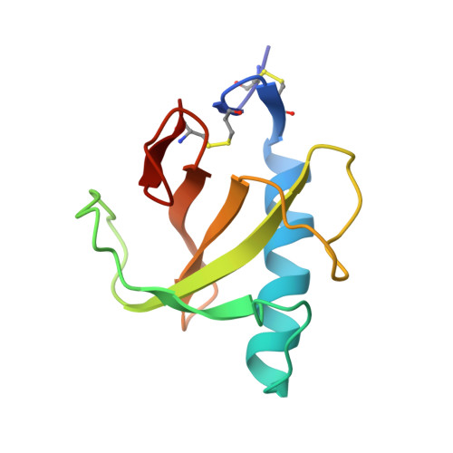
Three-dimensional structure of ribonuclease T1 complexed with guanylyl-2',5'-guanosine at 1.8 A resolution.
Koepke, J., Maslowska, M., Heinemann, U., Saenger, W.(1989) J Mol Biol 206: 475-488
- PubMed: 2541256
- DOI: https://doi.org/10.1016/0022-2836(89)90495-6
- Primary Citation of Related Structures:
2RNT - PubMed Abstract:
The enzyme ribonuclease T1 (RNase T1) isolated from Aspergillus oryzae was cocrystallized with the specific inhibitor guanylyl-2',5'-guanosine (2',5'-GpG) and the structure refined by the stereochemically restrained least-squares refinement method to a crystallographic R-factor of 14.9% for X-ray data above 3 sigma in the resolution range 6 to 1.8 A. The refined model consists of 781 protein atoms, 43 inhibitor atoms in a major site and 29 inhibitor atoms in a minor site, 107 water oxygen atoms, and a metal site assigned as Ca. At the end of the refinement, the orientation of His, Asn and Gln side-chains was reinterpreted on the basis of two-dimensional nuclear magnetic resonance data. The crystal packing and enzyme conformation of the RNase T1/2',5'-GpG complex and of the near-isomorphous RNase T1/2'-GMP complex are comparable. The root-mean-square deviation is 0.73 A between equivalent protein atoms. Differences in the unit cell dimensions are mainly due to the bound inhibitor. The 5'-terminal guanine of 2',5'-GpG binds to RNase T1 in much the same way as in the 2'-GMP complex. In contrast, the hydrogen bonds between the catalytic center and the phosphate group are different and the 3'-terminal guanine forms no hydrogen bonds with the enzyme. This poor binding is reflected in a 2-fold disorder of 2',5'-GpG (except the 5'-terminal guanine), which originates from differences in the pucker of the 5'-terminal ribose. The pucker is C2'-exo for the major site (2/3 occupancy) and C1'-endo for the minor site (1/3 occupancy). The orientation of the major site is stabilized through stacking interactions between the 3'-terminal guanine and His92, an amino acid necessary for catalysis. This might explain the high inhibition rate observed for 2',5'-GpG, which exceeds that of all other inhibitors of type 2',5'-GpN. On the basis of distance criteria, one solvent peak in the electron density was identified as metal ion, probably Ca2+. The ion is co-ordinated by the two Asp15 carboxylate oxygen atoms and by six water molecules. The co-ordination polyhedron displays approximate 4m2 symmetry.
Organizational Affiliation:
Institut für Kristallographie, Freie Universität Berlin, F.R.G.