2RMB

Crystal structures of cyclophilin A complexed with cyclosporin A and N-methyl-4-[(E)-2-butenyl]-4,4-dimethylthreonine cyclosporin A


Experimental Data Snapshot

  • Method: X-RAY DIFFRACTION
  • Resolution: 2.10 Å
  • R-Value Work: 0.184 
  • R-Value Observed: 0.184 

wwPDB Validation   3D Report Full Report


This is version 1.5 of the entry. See complete history


Literature

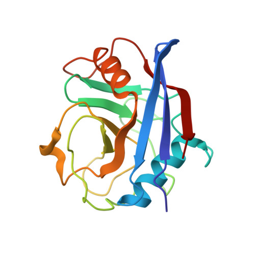
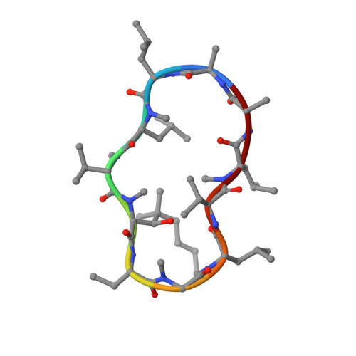
Crystal Structures of Cyclophilin a Complexed with Cyclosporin a and N-Methyl-4-[(E)-2-Butenyl]-4,4-Dimethylthreonine Cyclosporin A.

Ke, H.Mayrose, D.Belshaw, P.J.Alberg, D.G.Schreiber, S.L.Chang, Z.Y.Etzkorn, F.A.Ho, S.Walsh, C.T.

(1994) Structure 2: 33

  • DOI: https://doi.org/10.1016/s0969-2126(00)00006-x
  • Primary Citation of Related Structures:  
    2RMA, 2RMB

  • PubMed Abstract: 

    Cyclophilin (CyP) is a ubiquitious intracellular protein that binds the immunosuppressive drug cyclosporin A (CsA). CyP-CsA forms a ternary complex with calcineurin and thereby inhibits T-cell activation. CyP also has enzymatic activity, catalyzing the cis-trans isomerization of peptidyl-prolyl amide bonds. We have determined the structure of human cyclophilin A (CyPA) complexed with CsA to 2.1 A resolution. We also report here the structure of CyPA complexed with an analog of CsA, CsA (MeBm2t1-CsA), which binds less well to CyPA, but has increased immunosuppressive activity. Comparison of these structures with previously determined structures of unligated CyPA and CyPA complexed with a candidate substrate for the isomerase activity, the dipeptide AlaPro, reveals that subtle conformational changes occur in both CsA and CyPA on complex formation. MeBm2t1-CsA binds to CyPA in an essentially similar manner to CsA. The 100-fold weaker affinity of its binding may be attributable to the close contact between MeBmt1 and the active site residue Ala103 of CyPA, which causes small conformational changes in both protein and drug. One change, the slight movement of MeLeu6 in CsA relative to MeBm2t1-CsA, may be at least partially responsible for the higher affinity of the CyPA-MeBm2t1-CsA complex for calcineurin. Our comparison between CyPA-CsA and CyPA-AlaPro suggests that CsA is probably not an analog of the natural substrate, confirming that the catalytic activity of CyPA is not related to its role in immunosuppression either structurally or functionally.


  • Organizational Affiliation

    Department of Biochemistry and Biophysics, School of Medicine, University of North Carolina, Chapel HIll 27599.


Macromolecules
Find similar proteins by:  (by identity cutoff)  |  3D Structure
Entity ID: 1
MoleculeChains Sequence LengthOrganismDetailsImage
PEPTIDYL-PROLYL CIS-TRANS ISOMERASE
A, C, E, G, I
A, C, E, G, I, K, M, O, Q, S
165Homo sapiensMutation(s): 0 
Gene Names: CYCLOPHILIN A
EC: 5.2.1.8
UniProt & NIH Common Fund Data Resources
Find proteins for P62937 (Homo sapiens)
Explore P62937 
Go to UniProtKB:  P62937
PHAROS:  P62937
GTEx:  ENSG00000196262 
Entity Groups  
Sequence Clusters30% Identity50% Identity70% Identity90% Identity95% Identity100% Identity
UniProt GroupP62937
Sequence Annotations
Expand
  • Reference Sequence

Find similar proteins by:  Sequence   |   3D Structure  

Entity ID: 2
MoleculeChains Sequence LengthOrganismDetailsImage
CYCLOSPORIN A
B, D, F, H, J
B, D, F, H, J, L, N, P, R, T
11Tolypocladium inflatumMutation(s): 1 
Entity Groups  
Sequence Clusters30% Identity50% Identity70% Identity90% Identity95% Identity100% Identity
Sequence Annotations
Expand
  • Reference Sequence
Small Molecules
Modified Residues  4 Unique
IDChains TypeFormula2D DiagramParent
ABA
Query on ABA
B, D, F, H, J
B, D, F, H, J, L, N, P, R, T
L-PEPTIDE LINKINGC4 H9 N O2ALA
MLE
Query on MLE
B, D, F, H, J
B, D, F, H, J, L, N, P, R, T
L-PEPTIDE LINKINGC7 H15 N O2LEU
MVA
Query on MVA
B, D, F, H, J
B, D, F, H, J, L, N, P, R, T
L-PEPTIDE LINKINGC6 H13 N O2VAL
SAR
Query on SAR
B, D, F, H, J
B, D, F, H, J, L, N, P, R, T
PEPTIDE LINKINGC3 H7 N O2GLY
Biologically Interesting Molecules (External Reference) 1 Unique
Experimental Data & Validation

Experimental Data

  • Method: X-RAY DIFFRACTION
  • Resolution: 2.10 Å
  • R-Value Work: 0.184 
  • R-Value Observed: 0.184 
  • Space Group: P 1 21 1
Unit Cell:
Length ( Å )Angle ( ˚ )
a = 72.6α = 90
b = 160.6β = 90.9
c = 95.3γ = 90
Software Package:
Software NamePurpose
X-PLORmodel building
X-PLORrefinement
X-PLORphasing

Structure Validation

View Full Validation Report


Entry History 

Deposition Data

Revision History  (Full details and data files)

  • Version 1.0: 1995-02-07
    Type: Initial release
  • Version 1.1: 2011-06-14
    Changes: Version format compliance
  • Version 1.2: 2011-07-13
    Changes: Version format compliance
  • Version 1.3: 2011-07-27
    Changes: Atomic model, Database references, Derived calculations, Non-polymer description, Structure summary
  • Version 1.4: 2012-12-12
    Changes: Other
  • Version 1.5: 2017-11-01
    Changes: Derived calculations, Other