2R9Y

Structure of antiplasmin


Experimental Data Snapshot

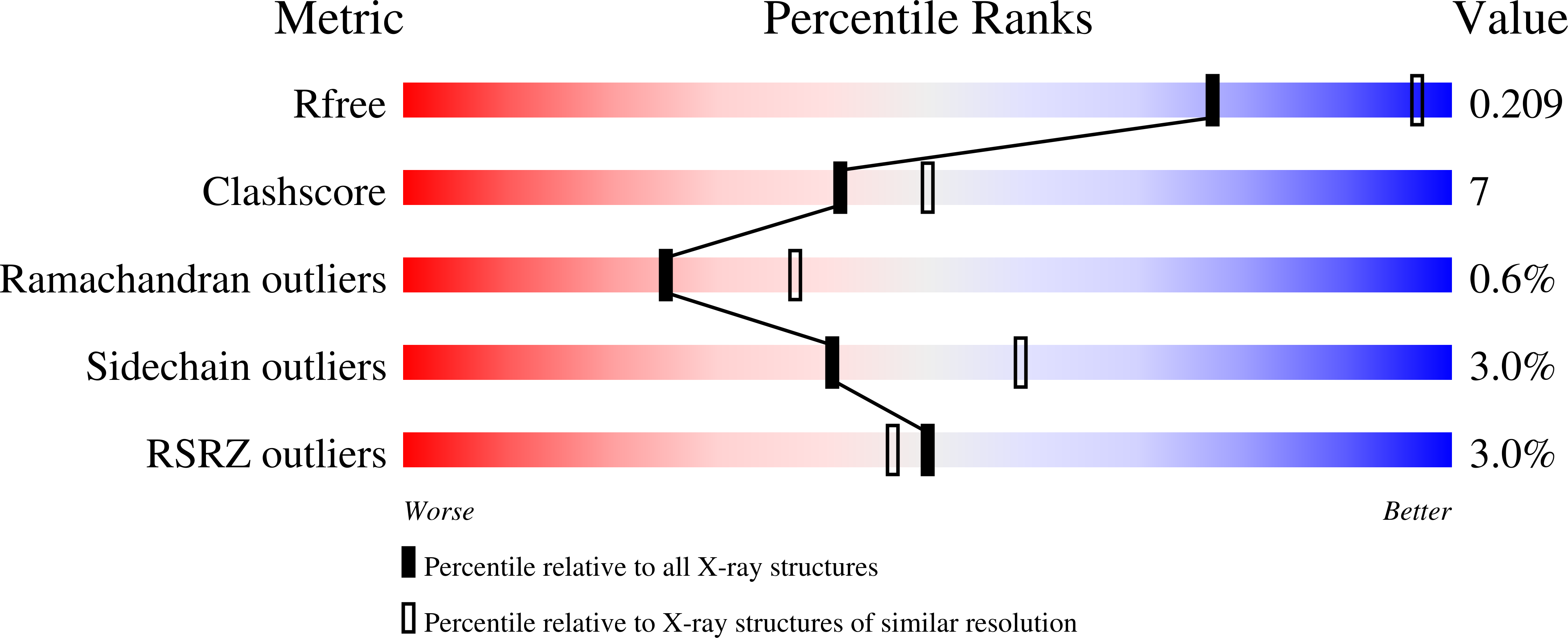
  • Method: X-RAY DIFFRACTION
  • Resolution: 2.65 Å
  • R-Value Free: 0.218 
  • R-Value Work: 0.184 
  • R-Value Observed: 0.186 

Starting Models: experimental
View more details

wwPDB Validation   3D Report Full Report


This is version 1.2 of the entry. See complete history


Literature

X-ray crystal structure of the fibrinolysis inhibitor {alpha}2-antiplasmin

Law, R.H.P.Sofian, T.Kan, W.T.Horvath, A.J.Hitchen, C.R.Langendorf, C.G.Buckle, A.M.Whisstock, J.C.Coughlin, P.B.

(2008) Blood 111: 2049-2052

  • DOI: https://doi.org/10.1182/blood-2007-09-114215
  • Primary Citation of Related Structures:  
    2R9Y

  • PubMed Abstract: 

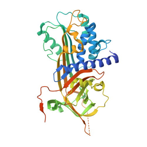
    The serpin alpha(2)-antiplasmin (SERPINF2) is the principal inhibitor of plasmin and inhibits fibrinolysis. Accordingly, alpha(2)-antiplasmin deficiency in humans results in uncontrolled fibrinolysis and a bleeding disorder. alpha(2)-antiplasmin is an unusual serpin, in that it contains extensive N- and C-terminal sequences flanking the serpin domain. The N-terminal sequence is crosslinked to fibrin by factor XIIIa, whereas the C-terminal region mediates the initial interaction with plasmin. To understand how this may happen, we have determined the 2.65A X-ray crystal structure of an N-terminal truncated murine alpha(2)-antiplasmin. The structure reveals that part of the C-terminal sequence is tightly associated with the body of the serpin. This would be anticipated to position the flexible plasmin-binding portion of the C-terminus in close proximity to the serpin Reactive Center Loop where it may act as a template to accelerate serpin/protease interactions.


  • Organizational Affiliation

    Department of Biochemistry and Molecular Biology, Monash University, Clayton, Australia


Macromolecules
Find similar proteins by:  (by identity cutoff)  |  3D Structure
Entity ID: 1
MoleculeChains Sequence LengthOrganismDetailsImage
Alpha-2-antiplasmin430Mus musculusMutation(s): 0 
Gene Names: Serpinf2Pli
UniProt
Find proteins for Q61247 (Mus musculus)
Explore Q61247 
Go to UniProtKB:  Q61247
Entity Groups  
Sequence Clusters30% Identity50% Identity70% Identity90% Identity95% Identity100% Identity
UniProt GroupQ61247
Sequence Annotations
Expand
  • Reference Sequence
Experimental Data & Validation

Experimental Data

  • Method: X-RAY DIFFRACTION
  • Resolution: 2.65 Å
  • R-Value Free: 0.218 
  • R-Value Work: 0.184 
  • R-Value Observed: 0.186 
  • Space Group: P 65
Unit Cell:
Length ( Å )Angle ( ˚ )
a = 115.679α = 90
b = 115.679β = 90
c = 100.533γ = 120
Software Package:
Software NamePurpose
REFMACrefinement
CrystalCleardata collection
MOSFLMdata reduction
SCALAdata scaling
PHASESphasing

Structure Validation

View Full Validation Report


Entry History 

Deposition Data

Revision History  (Full details and data files)

  • Version 1.0: 2007-12-18
    Type: Initial release
  • Version 1.1: 2011-07-13
    Changes: Advisory, Refinement description, Version format compliance
  • Version 1.2: 2023-10-25
    Changes: Data collection, Database references, Refinement description