2QK8

Crystal structure of the anthrax drug target, Bacillus anthracis dihydrofolate reductase


Experimental Data Snapshot

  • Method: X-RAY DIFFRACTION
  • Resolution: 2.40 Å
  • R-Value Free: 0.274 
  • R-Value Work: 0.243 
  • R-Value Observed: 0.244 

wwPDB Validation   3D Report Full Report


Ligand Structure Quality Assessment 


This is version 1.2 of the entry. See complete history


Literature

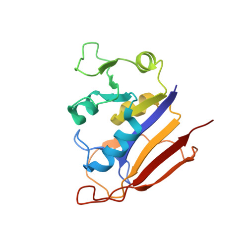
Crystal structure of the anthrax drug target, Bacillus anthracis dihydrofolate reductase.

Bennett, B.C.Xu, H.Simmerman, R.F.Lee, R.E.Dealwis, C.G.

(2007) J Med Chem 50: 4374-4381

  • DOI: https://doi.org/10.1021/jm070319v
  • Primary Citation of Related Structures:  
    2QK8

  • PubMed Abstract: 

    Spores of Bacillus anthracis are the infectious agent of anthrax. Current antibiotic treatments are limited due to resistance and patient age restrictions; thus, additional targets for therapeutic intervention are needed. One possible candidate is dihydrofolate reductase (DHFR), a biosynthetic enzyme necessary for anthrax pathogenicity. We determined the crystal structure of DHFR from B. anthracis (baDHFR) in complex with methotrexate (MTX; 1) at 2.4 Angstrom resolution. The structure reveals the crucial interactions required for MTX binding and a putative molecular basis for how baDHFR has natural resistance to trimethoprim (TMP; 2). The structure also allows insights for designing selective baDHFR inhibitors that will have weak affinities for the human enzyme. Additionally, we have found that 5-nitro-6-methylamino-isocytosine (MANIC; 3), which inhibits another B. anthracis folate synthesis enzyme, dihydropteroate synthase (DHPS), can also inhibit baDHFR. This provides a starting point for designing multi-target inhibitors that are less likely to induce drug resistance.


  • Organizational Affiliation

    Department of Biochemistry, Cellular and Molecular Biology, University of Tennessee, Knoxville, Tennessee 37996, USA.


Macromolecules
Find similar proteins by:  (by identity cutoff)  |  3D Structure
Entity ID: 1
MoleculeChains Sequence LengthOrganismDetailsImage
Dihydrofolate reductase162Bacillus anthracis str. SterneMutation(s): 0 
Gene Names: dfrA
EC: 1.5.1.3
UniProt
Find proteins for Q81R22 (Bacillus anthracis)
Explore Q81R22 
Go to UniProtKB:  Q81R22
Entity Groups  
Sequence Clusters30% Identity50% Identity70% Identity90% Identity95% Identity100% Identity
UniProt GroupQ81R22
Sequence Annotations
Expand
  • Reference Sequence
Small Molecules
Ligands 1 Unique
IDChains Name / Formula / InChI Key2D Diagram3D Interactions
MTX
Query on MTX

Download Ideal Coordinates CCD File 
B [auth A]METHOTREXATE
C20 H22 N8 O5
FBOZXECLQNJBKD-ZDUSSCGKSA-N
Binding Affinity Annotations 
IDSourceBinding Affinity
MTX BindingDB:  2QK8 IC50: min: 12.2, max: 24 (nM) from 4 assay(s)
Binding MOAD:  2QK8 IC50: 20 (nM) from 1 assay(s)
PDBBind:  2QK8 IC50: 12.2 (nM) from 1 assay(s)
Experimental Data & Validation

Experimental Data

  • Method: X-RAY DIFFRACTION
  • Resolution: 2.40 Å
  • R-Value Free: 0.274 
  • R-Value Work: 0.243 
  • R-Value Observed: 0.244 
  • Space Group: C 1 2 1
Unit Cell:
Length ( Å )Angle ( ˚ )
a = 82.697α = 90
b = 41.236β = 119.2
c = 67.032γ = 90
Software Package:
Software NamePurpose
REFMACrefinement
BioCARSdata collection
HKL-2000data reduction
HKL-2000data scaling
PHASERphasing

Structure Validation

View Full Validation Report


Ligand Structure Quality Assessment 


Entry History 

Deposition Data

Revision History  (Full details and data files)

  • Version 1.0: 2007-08-28
    Type: Initial release
  • Version 1.1: 2011-07-13
    Changes: Source and taxonomy, Version format compliance
  • Version 1.2: 2023-08-30
    Changes: Data collection, Database references, Refinement description