2QCA

A New Crystal Form of Bovine Pancreatic RNase A in Complex with 2'-Deoxyguanosine-5'-monophosphate


Experimental Data Snapshot

  • Method: X-RAY DIFFRACTION
  • Resolution: 1.33 Å
  • R-Value Free: 0.177 
  • R-Value Work: 0.119 
  • R-Value Observed: 0.125 

wwPDB Validation   3D Report Full Report


Ligand Structure Quality Assessment 


This is version 1.3 of the entry. See complete history


Literature

A new crystal form of bovine pancreatic RNase A in complex with 2'-deoxyguanosine-5'-monophosphate.

Larson, S.B.Day, J.S.Cudney, R.McPherson, A.

(2007) Acta Crystallogr Sect F Struct Biol Cryst Commun 63: 728-733

  • DOI: https://doi.org/10.1107/S1744309107039565
  • Primary Citation of Related Structures:  
    2QCA

  • PubMed Abstract: 

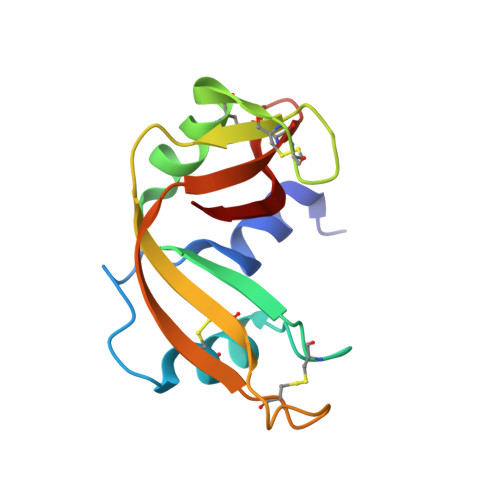
    The structure of bovine pancreatic RNase A has been determined in complex with 2'-deoxyguanosine-5'-monophosphate (dGMP) at 1.33 A resolution at room temperature in a previously unreported unit cell belonging to space group P3(1). There are two molecules of nucleotide per enzyme molecule, one of which lies in the active-site cleft in the productive binding mode, whilst the guanine base of the other dGMP occupies the pyrimidine-specific binding site in a nonproductive mode such that it forms hydrogen bonds to the phosphate group of the first dGMP. This is the first RNase A structure containing a guanine base in the B2 binding site. Each dGMP molecule is involved in intermolecular interactions with adjacent RNase A molecules in the lattice and the two nucleotides appear to direct the formation of the crystal lattice. Because GMP may be produced during degradation of RNA, this association could represent an inhibitor complex and thereby affect the observed enzyme kinetics.


  • Organizational Affiliation

    Department of Molecular Biology and Biochemistry, The University of California, Irvine, CA 92697-3900, USA.


Macromolecules
Find similar proteins by:  (by identity cutoff)  |  3D Structure
Entity ID: 1
MoleculeChains Sequence LengthOrganismDetailsImage
Ribonuclease pancreatic124Bos taurusMutation(s): 0 
EC: 3.1.27.5
UniProt
Find proteins for P61823 (Bos taurus)
Explore P61823 
Go to UniProtKB:  P61823
Entity Groups  
Sequence Clusters30% Identity50% Identity70% Identity90% Identity95% Identity100% Identity
UniProt GroupP61823
Sequence Annotations
Expand
  • Reference Sequence
Small Molecules
Ligands 1 Unique
IDChains Name / Formula / InChI Key2D Diagram3D Interactions
DGP
Query on DGP

Download Ideal Coordinates CCD File 
B [auth A],
C [auth A]
2'-DEOXYGUANOSINE-5'-MONOPHOSPHATE
C10 H14 N5 O7 P
LTFMZDNNPPEQNG-KVQBGUIXSA-N
Experimental Data & Validation

Experimental Data

  • Method: X-RAY DIFFRACTION
  • Resolution: 1.33 Å
  • R-Value Free: 0.177 
  • R-Value Work: 0.119 
  • R-Value Observed: 0.125 
  • Space Group: P 31
Unit Cell:
Length ( Å )Angle ( ˚ )
a = 55.08α = 90
b = 55.08β = 90
c = 39.2γ = 120
Software Package:
Software NamePurpose
SHELXmodel building
SHELXL-97refinement
d*TREKdata reduction
d*TREKdata scaling
CNSphasing

Structure Validation

View Full Validation Report


Ligand Structure Quality Assessment 


Entry History 

Deposition Data

Revision History  (Full details and data files)

  • Version 1.0: 2007-07-03
    Type: Initial release
  • Version 1.1: 2008-05-01
    Changes: Version format compliance
  • Version 1.2: 2011-07-13
    Changes: Non-polymer description, Version format compliance
  • Version 1.3: 2023-08-30
    Changes: Data collection, Database references, Derived calculations, Refinement description