Solution Structure of Inhibitor-Free Human Metalloelastase (MMP-12) Indicates an Internal Conformational Adjustment.
Bhaskaran, R., Palmier, M.O., Bagegni, N.A., Liang, X., Van Doren, S.R.(2007) J Mol Biol 374: 1333-1344
- PubMed: 17997411
- DOI: https://doi.org/10.1016/j.jmb.2007.10.028
- Primary Citation of Related Structures:
2POJ - PubMed Abstract:
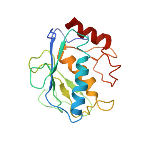
Macrophage metalloelastase or matrix metalloproteinase-12 (MMP-12) appears to exacerbate atherosclerosis, emphysema, aortic aneurysm, rheumatoid arthritis, and inflammatory bowel disease. An inactivating E219A mutation, validated by crystallography and NMR spectra, prevents autolysis of MMP-12 and allows us to determine its NMR structure without an inhibitor. The structural ensemble of the catalytic domain without an inhibitor is based on 2813 nuclear Overhauser effects (NOEs) and has an average RMSD to the mean structure of 0.25 A for the backbone and 0.61 A for all heavy atoms for residues Trp109-Gly263. Compared to crystal structures of MMP-12, helix B (hB) at the active site is unexpectedly more deeply recessed under the beta-sheet. This opens a pocket between hB and beta-strand IV in the active-site cleft. Both hB and an internal cavity are shifted toward beta-strand I, beta-strand III, and helix A on the back side of the protease. About 25 internal NOE contacts distinguish the inhibitor-free solution structure and indicate hB's greater depth and proximity to the sheet and helix A. Line broadening and multiplicity of amide proton NMR peaks from hB are consistent with hB undergoing a slow conformational exchange among subtly different environments. Inhibitor-binding-induced perturbations of the NMR spectra of MMP-1 and MMP-3 map to similar locations across MMP-12 and encompass the internal conformational adjustments. Evolutionary trace analysis suggests a functionally important network of residues that encompasses most of the locations adjusting in conformation, including 18 residues with NOE contacts unique to inhibitor-free MMP-12. The conformational change, sequence analysis, and inhibitor perturbations of NMR spectra agree on the network they identify between structural scaffold and the active site of MMPs.
Organizational Affiliation:
Department of Biochemistry, 117 Schweitzer Hall, University of Missouri, Columbia, MO 65211, USA.