2OK0

Fab ED10-DNA complex


Experimental Data Snapshot

  • Method: X-RAY DIFFRACTION
  • Resolution: 1.89 Å
  • R-Value Free: 0.204 
  • R-Value Work: 0.177 
  • R-Value Observed: 0.178 

wwPDB Validation   3D Report Full Report


This is version 1.3 of the entry. See complete history


Literature

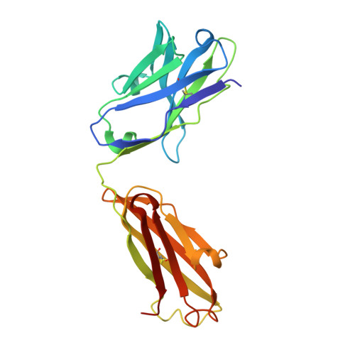
Specific recognition of a DNA immunogen by its elicited antibody

Sanguineti, S.Centeno Crowley, J.M.Lodeiro Merlo, M.F.Cerutti, M.L.Wilson, I.A.Goldbaum, F.A.Stanfield, R.L.de Prat-Gay, G.

(2007) J Mol Biol 370: 183-195

  • DOI: https://doi.org/10.1016/j.jmb.2007.04.046
  • Primary Citation of Related Structures:  
    2OJZ, 2OK0

  • PubMed Abstract: 

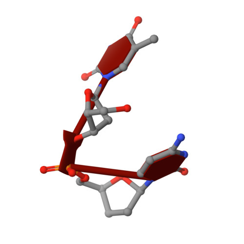
    DNA recognition by antibodies is a key feature of autoimmune diseases, yet model systems with structural information are very limited. The monoclonal antibody ED-10 recognizes one of the strands of the DNA duplex used in the immunogenic complex. Modifications of the 5' end decrease the binding affinity and short oligonucleotides retain high binding affinity. We determined crystal structures for the Fab bound to a 6-mer oligonucleotide containing the specific sequence that raised the antibody and compared it with the unliganded Fab. Only the first two bases from the 5' end (dTdC) display electron density and we observe four key hydrogen bonds at the interface. The thymine ring is stacked between TrpH50 and TrpH95, and the cytosine ring is packed against TyrL32. Upon DNA binding, TyrH97 and TrpH95 rearrange to allow subnanomolar binding affinity, five orders of magnitude higher than other reported complexes, possibly because of having gone through affinity maturation. This structure represents the first bona fide antibody DNA immunogen complex described in atomic detail.


  • Organizational Affiliation

    Instituto Leloir and CONICET, Patricias Argentinas 435, 1405 Buenos Aires, Argentina.


Macromolecules

Find similar proteins by:  (by identity cutoff)  |  3D Structure
Entity ID: 2
MoleculeChains Sequence LengthOrganismDetailsImage
Fab ED10 light chainB [auth L]219Mus musculusMutation(s): 0 
UniProt
Find proteins for A2NHM3 (Mus musculus)
Explore A2NHM3 
Go to UniProtKB:  A2NHM3
Entity Groups  
Sequence Clusters30% Identity50% Identity70% Identity90% Identity95% Identity100% Identity
UniProt GroupA2NHM3
Sequence Annotations
Expand
  • Reference Sequence
Find similar proteins by:  (by identity cutoff)  |  3D Structure
Entity ID: 3
MoleculeChains Sequence LengthOrganismDetailsImage
Fab ED10 heavy chainC [auth H]216Mus musculusMutation(s): 0 
UniProt
Find proteins for A0N7J2 (Mus sp)
Explore A0N7J2 
Go to UniProtKB:  A0N7J2
Entity Groups  
Sequence Clusters30% Identity50% Identity70% Identity90% Identity95% Identity100% Identity
UniProt GroupA0N7J2
Sequence Annotations
Expand
  • Reference Sequence

Find similar nucleic acids by:  Sequence   |   3D Structure  

Entity ID: 1
MoleculeChains LengthOrganismImage
5'-D(*TP*C)-3'A [auth D]2N/A
Sequence Annotations
Expand
  • Reference Sequence
Experimental Data & Validation

Experimental Data

  • Method: X-RAY DIFFRACTION
  • Resolution: 1.89 Å
  • R-Value Free: 0.204 
  • R-Value Work: 0.177 
  • R-Value Observed: 0.178 
  • Space Group: C 1 2 1
Unit Cell:
Length ( Å )Angle ( ˚ )
a = 123.711α = 90
b = 63.833β = 116.26
c = 83.88γ = 90
Software Package:
Software NamePurpose
REFMACrefinement
DENZOdata reduction
SCALEPACKdata scaling
EPMRphasing

Structure Validation

View Full Validation Report


Entry History 

Deposition Data

Revision History  (Full details and data files)

  • Version 1.0: 2007-06-12
    Type: Initial release
  • Version 1.1: 2008-05-01
    Changes: Version format compliance
  • Version 1.2: 2011-07-13
    Changes: Advisory, Version format compliance
  • Version 1.3: 2023-12-27
    Changes: Data collection, Database references