2OCJ

Human p53 core domain in the absence of DNA


Experimental Data Snapshot

  • Method: X-RAY DIFFRACTION
  • Resolution: 2.05 Å
  • R-Value Free: 0.246 
  • R-Value Work: 0.209 

wwPDB Validation   3D Report Full Report


This is version 1.4 of the entry. See complete history


Literature

Structure of the human p53 core domain in the absence of DNA.

Wang, Y.Rosengarth, A.Luecke, H.

(2007) Acta Crystallogr D Biol Crystallogr 63: 276-281

  • DOI: https://doi.org/10.1107/S0907444906048499
  • Primary Citation of Related Structures:  
    2OCJ

  • PubMed Abstract: 

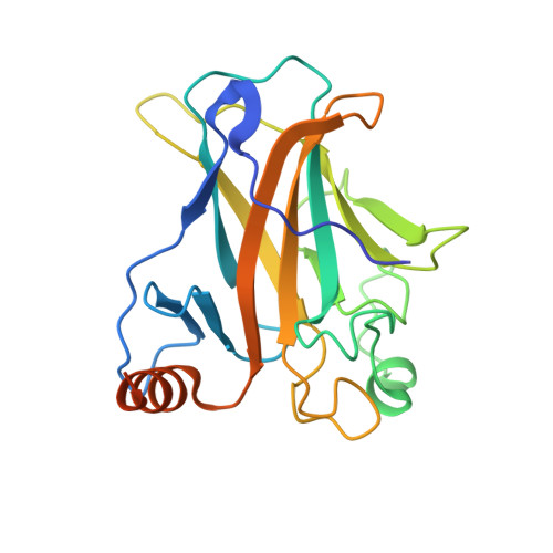
    The tumor suppressor protein p53 plays a key role in cell-cycle regulation by triggering DNA repair, cell-cycle arrest and apoptosis when the appropriate signal is received. p53 has the classic architecture of a transcription factor, with an amino-terminal transactivation domain, a core DNA-binding domain and carboxy-terminal tetramerization and regulatory domains. The crystal structure of the p53 core domain, which includes the amino acids from residue 96 to residue 289, has been determined in the absence of DNA to a resolution of 2.05 A. Crystals grew in a new monoclinic space group (P2(1)), with unit-cell parameters a = 68.91, b = 69.36, c = 84.18 A, beta = 90.11 degrees . The structure was solved by molecular replacement and has been refined to a final R factor of 20.9% (R(free) = 24.6%). The final model contains four molecules in the asymmetric unit with four zinc ions and 389 water molecules. The non-crystallographic tetramers display different protein contacts from those in other p53 crystals, giving rise to the question of how p53 arranges as a tetramer when it binds its target DNA.


  • Organizational Affiliation

    Department of Molecular Biology and Biochemistry, 3205 McGaugh Hall, University of California, Irvine, CA 92697-3900, USA.


Macromolecules
Find similar proteins by:  (by identity cutoff)  |  3D Structure
Entity ID: 1
MoleculeChains Sequence LengthOrganismDetailsImage
P53 TUMOR SUPPRESSOR
A, B, C, D
219Homo sapiensMutation(s): 0 
UniProt & NIH Common Fund Data Resources
Find proteins for P04637 (Homo sapiens)
Explore P04637 
Go to UniProtKB:  P04637
PHAROS:  P04637
GTEx:  ENSG00000141510 
Entity Groups  
Sequence Clusters30% Identity50% Identity70% Identity90% Identity95% Identity100% Identity
UniProt GroupP04637
Sequence Annotations
Expand
  • Reference Sequence
Experimental Data & Validation

Experimental Data

  • Method: X-RAY DIFFRACTION
  • Resolution: 2.05 Å
  • R-Value Free: 0.246 
  • R-Value Work: 0.209 
  • Space Group: P 1 21 1
Unit Cell:
Length ( Å )Angle ( ˚ )
a = 68.913α = 90
b = 69.364β = 90.11
c = 84.179γ = 90
Software Package:
Software NamePurpose
DENZOdata reduction
SCALEPACKdata scaling
MOLREPphasing
CNSrefinement
PDB_EXTRACTdata extraction
HKL-2000data collection
HKL-2000data reduction
PHASERphasing

Structure Validation

View Full Validation Report


Entry History 

Deposition Data

Revision History  (Full details and data files)

  • Version 1.0: 2007-03-06
    Type: Initial release
  • Version 1.1: 2008-05-01
    Changes: Version format compliance
  • Version 1.2: 2011-07-13
    Changes: Version format compliance
  • Version 1.3: 2017-10-18
    Changes: Refinement description
  • Version 1.4: 2023-08-30
    Changes: Data collection, Database references, Derived calculations, Refinement description