The short-lived signaling state of the photoactive yellow protein photoreceptor revealed by combined structural probes.
Ramachandran, P.L., Lovett, J.E., Carl, P.J., Cammarata, M., Lee, J.H., Jung, Y.O., Ihee, H., Timmel, C.R., van Thor, J.J.(2011) J Am Chem Soc 133: 9395-9404
- PubMed: 21627157
- DOI: https://doi.org/10.1021/ja200617t
- Primary Citation of Related Structures:
2KX6 - PubMed Abstract:
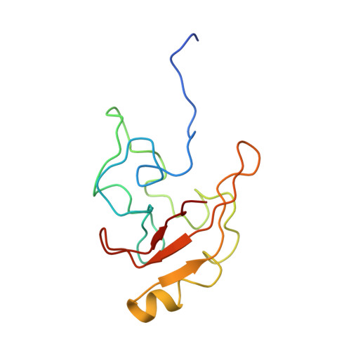
The signaling state of the photoactive yellow protein (PYP) photoreceptor is transiently developed via isomerization of its blue-light-absorbing chromophore. The associated structural rearrangements have large amplitude but, due to its transient nature and chemical exchange reactions that complicate NMR detection, its accurate three-dimensional structure in solution has been elusive. Here we report on direct structural observation of the transient signaling state by combining double electron electron resonance spectroscopy (DEER), NMR, and time-resolved pump-probe X-ray solution scattering (TR-SAXS/WAXS). Measurement of distance distributions for doubly spin-labeled photoreceptor constructs using DEER spectroscopy suggests that the signaling state is well ordered and shows that interspin-label distances change reversibly up to 19 Å upon illumination. The SAXS/WAXS difference signal for the signaling state relative to the ground state indicates the transient formation of an ordered and rearranged conformation, which has an increased radius of gyration, an increased maximum dimension, and a reduced excluded volume. Dynamical annealing calculations using the DEER derived long-range distance restraints in combination with short-range distance information from (1)H-(15)N HSQC perturbation spectroscopy give strong indication for a rearrangement that places part of the N-terminal domain in contact with the exposed chromophore binding cleft while the terminal residues extend away from the core. Time-resolved global structural information from pump-probe TR-SAXS/WAXS data supports this conformation and allows subsequent structural refinement that includes the combined energy terms from DEER, NMR, and SAXS/WAXS together. The resulting ensemble simultaneously satisfies all restraints, and the inclusion of TR-SAXS/WAXS effectively reduces the uncertainty arising from the possible spin-label orientations. The observations are essentially compatible with reduced folding of the I(2)' state (also referred to as the 'pB' state) that is widely reported, but indicates it to be relatively ordered and rearranged. Furthermore, there is direct evidence for the repositioning of the N-terminal region in the I(2)' state, which is structurally modeled by dynamical annealing and refinement calculations.
Organizational Affiliation:
Laboratory of Molecular Biophysics, Department of Biochemistry, University of Oxford, South Parks Road, Oxford OX1 3QU, UK.