Structural role of tyrosine 98 in photoactive yellow protein: effects on fluorescence, gateway, and photocycle recovery.
Kyndt, J.A., Savvides, S.N., Memmi, S., Koh, M., Fitch, J.C., Meyer, T.E., Heyn, M.P., Van Beeumen, J.J., Cusanovich, M.A.(2007) Biochemistry 46: 95-105
- PubMed: 17198379
- DOI: https://doi.org/10.1021/bi061867y
- Primary Citation of Related Structures:
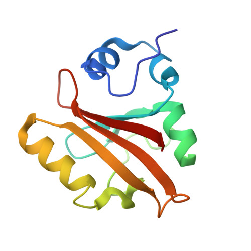
2I9V - PubMed Abstract:
We have recently shown that the Y98Q mutant of PYP has a major effect on the photocycle kinetics ( approximately 40 times slower recovery). We have now determined the crystal structure of Y98Q at 2.2 A resolution to reveal the role of residue Y98 in the PYP photocycle. Although the overall structure is very similar to that of WT, we observed two major effects of the mutation. One obvious consequence is a conformational change of the beta4-beta5 loop, which includes a repositioning of residue M100. It had previously been shown that the photocycle is slowed by as much as 3 orders of magnitude when residue M100 is substituted or when the conformation is altered as in Rhodocista centenaria PYP. To investigate whether the altered photocycle of Y98Q is due to this repositioning of M100 or is caused by an effect unrelated to M100, we determined the dark recovery kinetics of the Y98Q/M100A mutant. We find the recovery kinetics to be very similar to the M100A single mutant kinetics and therefore conclude that the slower recovery kinetics in Y98Q are most likely due to repositioning of M100. In addition, we find that other substitutions at position 98 (Y98W, Y98L, and Y98A) have differing effects on the photocycle recovery, presumably due to a variable distortion of the beta4-beta5 loop. The second effect of the Y98Q mutation is a repositioning of R52, which is thought to interact with Y98 in WT PYP and now forms new interactions with residues Q99 and Q56. To determine the role of R52, we also characterized an R52A/M100A double mutant and found that the effects on the recovery kinetics ( approximately 2000 slower recovery than WT) are due to unrelated events in the photocycle. Since the Y98Q/M100A recovery kinetics are more similar to those of M100 than R52A/M100A, we conclude that the repositioning of R52, caused by the Y98Q mutation, does not affect the dark state recovery. In addition, it has been proposed that Y98 and P68 are "gateway residues" between which the chromophore must pass during isomerization. We tested the recovery kinetics of mutant P68A and found that, although the gateway may be important for photocycle initiation, its role in recovery to the ground state is minimal.
Organizational Affiliation:
Department of Biochemistry and Molecular Biophysics, University of Arizona, Tucson, Arizona 85721, USA.