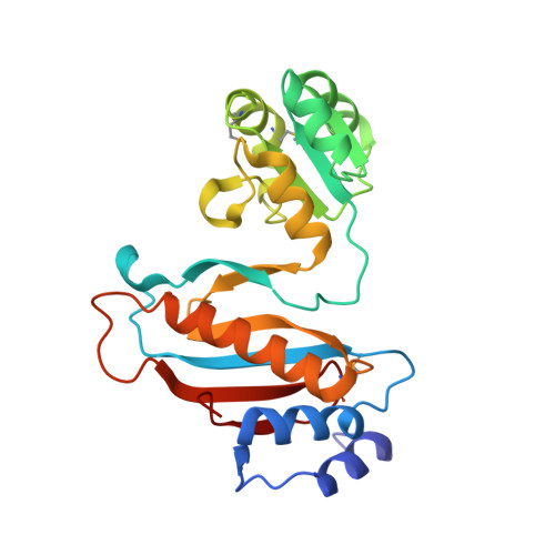
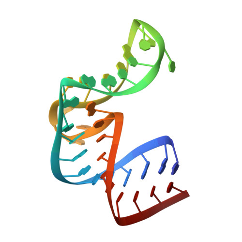
2HW8

Structure of ribosomal protein L1-mRNA complex at 2.1 resolution.


Experimental Data Snapshot

  • Method: X-RAY DIFFRACTION
  • Resolution: 2.10 Å
  • R-Value Free: 0.247 
  • R-Value Work: 0.212 
  • R-Value Observed: 0.212 

wwPDB Validation   3D Report Full Report


This is version 1.4 of the entry. See complete history


Literature

Structure of the ribosomal protein L1-mRNA complex at 2.1 A resolution: common features of crystal packing of L1-RNA complexes.

Tishchenko, S.Nikonova, E.Nikulin, A.Nevskaya, N.Volchkov, S.Piendl, W.Garber, M.Nikonov, S.

(2006) Acta Crystallogr D Biol Crystallogr 62: 1545-1554

  • DOI: https://doi.org/10.1107/S0907444906041655
  • Primary Citation of Related Structures:  
    2HW8

  • PubMed Abstract: 

    The crystal structure of a hybrid complex between the bacterial ribosomal protein L1 from Thermus thermophilus and a Methanococcus vannielii mRNA fragment containing an L1-binding site was determined at 2.1 A resolution. It was found that all polar atoms involved in conserved protein-RNA hydrogen bonds have high values of density in the electron-density map and that their hydrogen-bonding capacity is fully realised through interactions with protein atoms, water molecules and K(+) ions. Intermolecular contacts were thoroughly analyzed in the present crystals and in crystals of previously determined L1-RNA complexes. It was shown that extension of the RNA helices providing canonical helix stacking between open-open or open-closed ends of RNA fragments is a common feature of these and all known crystals of complexes between ribosomal proteins and RNAs. In addition, the overwhelming majority of complexes between ribosomal proteins and RNA molecules display crystal contacts formed by the central parts of the RNA fragments. These contacts are often very extensive and strong and it is proposed that they are formed in the saturated solution prior to crystal formation.


  • Organizational Affiliation

    Institute of Protein Research, Russian Academy of Sciences, 142290 Pushchino, Moscow Region, Russia.


Macromolecules

Find similar proteins by:  (by identity cutoff)  |  3D Structure
Entity ID: 2
MoleculeChains Sequence LengthOrganismDetailsImage
50S ribosomal protein L1B [auth A]228Thermus thermophilusMutation(s): 2 
Gene Names: rpl1
UniProt
Find proteins for P27150 (Thermus thermophilus)
Explore P27150 
Go to UniProtKB:  P27150
Entity Groups  
Sequence Clusters30% Identity50% Identity70% Identity90% Identity95% Identity100% Identity
UniProt GroupP27150
Sequence Annotations
Expand
  • Reference Sequence
Find similar nucleic acids by:  (by identity cutoff)  |  3D Structure
Entity ID: 1
MoleculeChains LengthOrganismImage
36-MERA [auth B]36N/A
Sequence Annotations
Expand
  • Reference Sequence
Small Molecules
Modified Residues  1 Unique
IDChains TypeFormula2D DiagramParent
MSE
Query on MSE
B [auth A]L-PEPTIDE LINKINGC5 H11 N O2 SeMET
Experimental Data & Validation

Experimental Data

  • Method: X-RAY DIFFRACTION
  • Resolution: 2.10 Å
  • R-Value Free: 0.247 
  • R-Value Work: 0.212 
  • R-Value Observed: 0.212 
  • Space Group: P 65 2 2
Unit Cell:
Length ( Å )Angle ( ˚ )
a = 67.87α = 90
b = 67.87β = 90
c = 340.47γ = 120
Software Package:
Software NamePurpose
CNSrefinement
XDSdata reduction
XSCALEdata scaling
CNSphasing

Structure Validation

View Full Validation Report


Entry History 

Deposition Data

Revision History  (Full details and data files)

  • Version 1.0: 2006-12-19
    Type: Initial release
  • Version 1.1: 2008-05-01
    Changes: Version format compliance
  • Version 1.2: 2011-07-13
    Changes: Version format compliance
  • Version 1.3: 2023-08-30
    Changes: Data collection, Database references, Derived calculations, Refinement description
  • Version 1.4: 2023-11-15
    Changes: Data collection