2H6Z

Crystal Structure of Thioredoxin Mutant E44D in Hexagonal (p61) Space Group


Experimental Data Snapshot

  • Method: X-RAY DIFFRACTION
  • Resolution: 2.25 Å
  • R-Value Free: 0.290 
  • R-Value Work: 0.228 
  • R-Value Observed: 0.234 

wwPDB Validation   3D Report Full Report


This is version 1.6 of the entry. See complete history


Literature

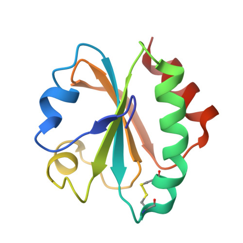
A stability pattern of protein hydrophobic mutations that reflects evolutionary structural optimization.

Godoy-Ruiz, R.Perez-Jimenez, R.Ibarra-Molero, B.Sanchez-Ruiz, J.M.

(2005) Biophys J 89: 3320-3331

  • DOI: https://doi.org/10.1529/biophysj.105.067025
  • Primary Citation of Related Structures:  
    2H6Z, 2H70

  • PubMed Abstract: 

    We have determined the effect of mutations involving isoleucine and valine (i.e., mutations I-->V and V-->I) on the stability of Escherichia coli thioredoxin. Despite the similarity in chemical structure (V and I differ only in a methyl group), we find that many environments are optimized to a significant extent for either V or I. We find, furthermore, that a plot of effect of hydrophobic mutations on stability versus packing density shows a strikingly simple pattern that clearly reflects evolutionary structural optimization. The existence of such patterns suggests the possibility of rationalizing (and perhaps even predicting) mutation effects on protein stability on the basis of evolutionary models. By "evolutionary model" we specifically refer in this context to a model for mutation effects on stability in which certain physical features of the mutated residue environments are evaluated from an assumption regarding how such environments have been selected during protein evolution (as opposed to a purely "physical model" in which those features would be derived from some kind of energetics analysis of the protein structural characteristics). To illustrate this novel approach and provide general guidelines for its application, we develop here a simple evolutionary model that successfully explains the effect of the I<-->V mutations on thioredoxin stability.


  • Organizational Affiliation

    Departamento de Quimica Fisica, Facultad de Ciencias, Universidad de Granada, Campus Fuentenueva s/n, 18071 Granada, Spain.


Macromolecules
Find similar proteins by:  (by identity cutoff)  |  3D Structure
Entity ID: 1
MoleculeChains Sequence LengthOrganismDetailsImage
Thioredoxin
A, B
108Escherichia coliMutation(s): 1 
Gene Names: trxA
UniProt
Find proteins for P0AA25 (Escherichia coli (strain K12))
Explore P0AA25 
Go to UniProtKB:  P0AA25
Entity Groups  
Sequence Clusters30% Identity50% Identity70% Identity90% Identity95% Identity100% Identity
UniProt GroupP0AA25
Sequence Annotations
Expand
  • Reference Sequence
Experimental Data & Validation

Experimental Data

  • Method: X-RAY DIFFRACTION
  • Resolution: 2.25 Å
  • R-Value Free: 0.290 
  • R-Value Work: 0.228 
  • R-Value Observed: 0.234 
  • Space Group: P 61
Unit Cell:
Length ( Å )Angle ( ˚ )
a = 102.916α = 90
b = 102.916β = 90
c = 42.559γ = 120
Software Package:
Software NamePurpose
SAINTdata scaling
MOLREPphasing
REFMACrefinement
PDB_EXTRACTdata extraction
PROTEUM PLUSdata reduction
SADABSdata scaling
XPREPdata reduction
Cootmodel building
MolProbitymodel building

Structure Validation

View Full Validation Report


Entry History 

Deposition Data

Revision History  (Full details and data files)

  • Version 1.0: 2007-05-15
    Type: Initial release
  • Version 1.1: 2008-05-01
    Changes: Version format compliance
  • Version 1.2: 2011-07-13
    Changes: Advisory, Version format compliance
  • Version 1.3: 2012-05-09
    Changes: Other
  • Version 1.4: 2017-10-18
    Changes: Refinement description
  • Version 1.5: 2021-10-20
    Changes: Database references, Derived calculations, Experimental preparation
  • Version 1.6: 2023-08-30
    Changes: Data collection, Refinement description