


Crystal Structure of Thioredoxin Mutant P34H
Gavira, J.A., Perez-Jimenez, R., Ibarra-Molero, B., Sanchez-Ruiz, J.M.To be published.
Experimental Data Snapshot
wwPDB Validation 3D Report Full Report

Entity ID: 1 | |||||
|---|---|---|---|---|---|
| Molecule | Chains | Sequence Length | Organism | Details | Image |
| Thioredoxin 1 | 108 | Escherichia coli | Mutation(s): 1 Gene Names: trxA, fipA, tsnC | 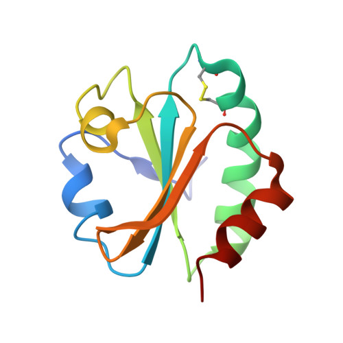 | |
UniProt | |||||
Find proteins for P0AA25 (Escherichia coli (strain K12)) Explore P0AA25 Go to UniProtKB: P0AA25 | |||||
Entity Groups | |||||
| Sequence Clusters | 30% Identity50% Identity70% Identity90% Identity95% Identity100% Identity | ||||
| UniProt Group | P0AA25 | ||||
Sequence AnnotationsExpand | |||||
| |||||
| Length ( Å ) | Angle ( ˚ ) |
|---|---|
| a = 31.138 | α = 90 |
| b = 90.041 | β = 112.99 |
| c = 36.326 | γ = 90 |
| Software Name | Purpose |
|---|---|
| REFMAC | refinement |
| PROTEUM PLUS | data reduction |
| SAINT | data scaling |
| SADABS | data scaling |
| XPREP | data reduction |
| MOLREP | phasing |
RCSB PDB (citation) is hosted by
RCSB PDB is a member of the

