The Stacking Tryptophan of Galactose Oxidase: A Second-Coordination Sphere Residue that Has Profound Effects on Tyrosyl Radical Behavior and Enzyme Catalysis
Rogers, M.S., Tyler, E.M., Akyumani, N., Kurtis, C.R., Spooner, R.K., Deacon, S.E., Tamber, S., Firbank, S.J., Mahmoud, K., Knowles, P.F., Phillips, S.E., McPherson, M.J., Dooley, D.M.(2007) Biochemistry 46: 4606-4618
- PubMed: 17385891
- DOI: https://doi.org/10.1021/bi062139d
- Primary Citation of Related Structures:
2EIB, 2EIC, 2EID, 2EIE - PubMed Abstract:
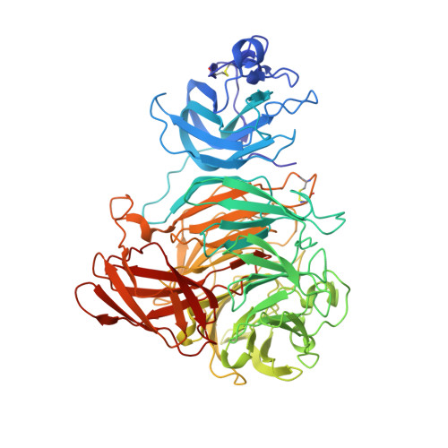
The function of the stacking tryptophan, W290, a second-coordination sphere residue in galactose oxidase, has been investigated via steady-state kinetics measurements, absorption, CD and EPR spectroscopy, and X-ray crystallography of the W290F, W290G, and W290H variants. Enzymatic turnover is significantly slower in the W290 variants. The Km for D-galactose for W290H is similar to that of the wild type, whereas the Km is greatly elevated in W290G and W290F, suggesting a role for W290 in substrate binding and/or positioning via the NH group of the indole ring. Hydrogen bonding between W290 and azide in the wild type-azide crystal structure are consistent with this function. W290 modulates the properties and reactivity of the redox-active tyrosine radical; the Y272 tyrosyl radicals in both the W290G and W290H variants have elevated redox potentials and are highly unstable compared to the radical in W290F, which has properties similar to those of the wild-type tyrosyl radical. W290 restricts the accessibility of the Y272 radical site to solvent. Crystal structures show that Y272 is significantly more solvent exposed in the W290G variant but that W290F limits solvent access comparable to the wild-type indole side chain. Spectroscopic studies indicate that the Cu(II) ground states in the semireduced W290 variants are very similar to that of the wild-type protein. In addition, the electronic structures of W290X-azide complexes are also closely similar to the wild-type electronic structure. Azide binding and azide-mediated proton uptake by Y495 are perturbed in the variants, indicating that tryptophan also modulates the function of the catalytic base (Y495) in the wild-type enzyme. Thus, W290 plays multiple critical roles in enzyme catalysis, affecting substrate binding, the tyrosyl radical redox potential and stability, and the axial tyrosine function.
Organizational Affiliation:
Department of Chemistry and Biochemistry, Montana State University, Bozeman, Montana 59717, USA.


