2CHE

STRUCTURE OF THE MG2+-BOUND FORM OF CHEY AND MECHANISM OF PHOSPHORYL TRANSFER IN BACTERIAL CHEMOTAXIS


Experimental Data Snapshot

  • Method: X-RAY DIFFRACTION
  • Resolution: 1.80 Å
  • R-Value Observed: 0.190 

wwPDB Validation   3D Report Full Report


This is version 1.4 of the entry. See complete history


Literature

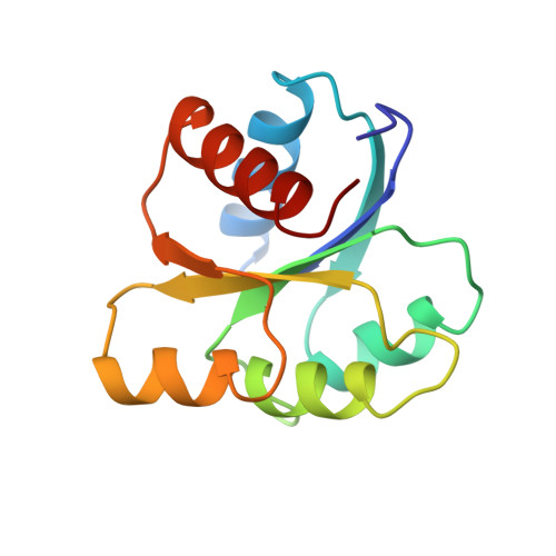
Structure of the Mg(2+)-bound form of CheY and mechanism of phosphoryl transfer in bacterial chemotaxis.

Stock, A.M.Martinez-Hackert, E.Rasmussen, B.F.West, A.H.Stock, J.B.Ringe, D.Petsko, G.A.

(1993) Biochemistry 32: 13375-13380

  • DOI: https://doi.org/10.1021/bi00212a001
  • Primary Citation of Related Structures:  
    2CHE, 2CHF

  • PubMed Abstract: 

    The response regulator protein of bacterial chemotaxis, CheY, is representative of a large family of signal transduction proteins that function as phosphorylation-activated switches to regulate the activities of associated effector domains. These regulators catalyze the metal ion-dependent phosphoryl transfer and dephosphorylation reactions that control the effector activities. The crystal structures of Salmonella typhimurium CheY with and without Mg2+ bound at the active site have been determined and refined at 1.8-A resolution. While the overall structures of metal-bound and metal-free CheY are similar, significant rearrangements occur within the active site involving the three most highly conserved residues of the response regulator family. Conservation of the cluster of carboxylate side chains at the active site of response regulator domains can be rationalized in terms of their role in coordinating the catalytically essential divalent metal ion. The Mg2+ coordination geometry provides insights to the mechanism of phosphoryl transfer.


  • Organizational Affiliation

    Center for Advanced Biotechnology and Medicine, Piscataway, New Jersey.


Macromolecules
Find similar proteins by:  (by identity cutoff)  |  3D Structure
Entity ID: 1
MoleculeChains Sequence LengthOrganismDetailsImage
CHEY128Salmonella enterica subsp. enterica serovar TyphimuriumMutation(s): 0 
UniProt
Find proteins for P0A2D5 (Salmonella typhimurium (strain LT2 / SGSC1412 / ATCC 700720))
Explore P0A2D5 
Go to UniProtKB:  P0A2D5
Entity Groups  
Sequence Clusters30% Identity50% Identity70% Identity90% Identity95% Identity100% Identity
UniProt GroupP0A2D5
Sequence Annotations
Expand
  • Reference Sequence
Small Molecules
Ligands 1 Unique
IDChains Name / Formula / InChI Key2D Diagram3D Interactions
MG
Query on MG

Download Ideal Coordinates CCD File 
B [auth A]MAGNESIUM ION
Mg
JLVVSXFLKOJNIY-UHFFFAOYSA-N
Experimental Data & Validation

Experimental Data

  • Method: X-RAY DIFFRACTION
  • Resolution: 1.80 Å
  • R-Value Observed: 0.190 
  • Space Group: P 21 21 21
Unit Cell:
Length ( Å )Angle ( ˚ )
a = 45.71α = 90
b = 46.98β = 90
c = 53.91γ = 90
Software Package:
Software NamePurpose
PROLSQrefinement

Structure Validation

View Full Validation Report


Entry History 

Deposition Data

Revision History  (Full details and data files)

  • Version 1.0: 1994-04-30
    Type: Initial release
  • Version 1.1: 2008-03-03
    Changes: Version format compliance
  • Version 1.2: 2011-07-13
    Changes: Version format compliance
  • Version 1.3: 2017-11-29
    Changes: Derived calculations, Other
  • Version 1.4: 2024-02-14
    Changes: Data collection, Database references, Derived calculations