Solution Structure of an Oncogenic Mutant of Cdc42Hs
Adams, P.D., Oswald, R.E.(2006) Biochemistry 45: 2577-2583
- PubMed: 16489751
- DOI: https://doi.org/10.1021/bi051686g
- Primary Citation of Related Structures:
2ASE - PubMed Abstract:
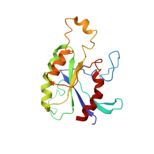
Cdc42Hs(F28L) is a single-point mutant of Cdc42Hs, a member of the Ras superfamily of GTP-binding proteins, that facilitates cellular transformation brought about by an increased rate of cycling between GTP and GDP [Lin, R., et al. (1997) Curr. Biol. 7, 794-797]. Dynamics studies of Cdc42Hs(F28L)-GDP have shown increased flexibility for several residues at the nucleotide-binding site [Adams, P. D., et al. (2004) Biochemistry 43, 9968-9977]. The solution structure of Cdc42Hs-GDP (wild type) has previously been determined by NMR spectroscopy [Feltham, J. L., et al. (1997) Biochemistry 36, 8755-8766]. Here, we describe the solution structure of Cdc42Hs(F28L)-GDP, which provides insight into the structural basis for the change in affinity for GDP. Heteronuclear NMR experiments were performed to assign resonances in the protein, and distance, hydrogen bonding, residual dipolar coupling, and dihedral angle constraints were used to calculate a set of low-energy structures using distance geometry and simulated annealing refinement protocols. The overall structure of Cdc42Hs(F28L)-GDP is very similar to that of wild-type Cdc42Hs, consisting of a centrally located six-stranded beta-sheet structure surrounding the C-terminal alpha-helix [Feltham, J. L., et al. (1997) Biochemistry 36, 8755-8766]. In addition, the same three regions in wild-type Cdc42Hs that show structural disorder (Switch I, Switch II, and the Insert region) are disordered in F28L as well. Although the structure of Cdc42Hs(F28L)-GDP is very similar to that of the wild type, interactions with the nucleotide and hydrogen bonding within the nucleotide binding site are altered, and the region surrounding L28 is substantially more disordered.
Organizational Affiliation:
Department of Molecular Medicine, Cornell University, Ithaca, New York 14853, USA.