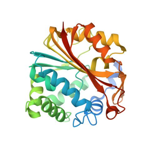
Mode of binding of methyl acceptor substrates to the adrenaline-synthesizing enzyme phenylethanolamine N-methyltransferase: implications for catalysis
Gee, C.L., Tyndall, J.D.A., Grunewald, G.L., Wu, Q., McLeish, M.J., Martin, J.L.(2005) Biochemistry 44: 16875-16885
- PubMed: 16363801
- DOI: https://doi.org/10.1021/bi051636b
- Primary Citation of Related Structures:
2AN3, 2AN4, 2AN5 - PubMed Abstract:
Here we report three crystal structure complexes of human phenylethanolamine N-methyltransferase (PNMT), one bound with a substrate that incorporates a flexible ethanolamine side chain (p-octopamine), a second bound with a semirigid analogue substrate [cis-(1R,2S)-2-amino-1-tetralol, cis-(1R,2S)-AT], and a third with trans-(1S,2S)-2-amino-1-tetralol [trans-(1S,2S)-AT] that acts as an inhibitor of PNMT rather than a substrate. A water-mediated interaction between the critical beta-hydroxyl of the flexible ethanolamine group of p-octopamine and an acidic residue, Asp267, is likely to play a key role in positioning the side chain correctly for methylation to occur at the amine. A second interaction with Glu219 may play a lesser role. Catalysis likely occurs via deprotonation of the amine through the action of Glu185; mutation of this residue significantly reduced the kcat without affecting the Km. The mode of binding of cis-(1R,2S)-AT supports the notion that this substrate is a conformationally restrained analogue of flexible PNMT substrates, in that it forms interactions with the enzyme similar to those observed for p-octopamine. By contrast, trans-(1S,2S)-AT, an inhibitor rather than a substrate, binds in an orientation that is flipped by 180 degrees compared with cis-(1R,2S)-AT. A consequence of this flipped binding mode is that the interactions between the hydroxyl and Asp267 and Glu219 are lost. However, the amines of inhibitor trans-(1S,2S)-AT and substrate cis-(1R,2S)-AT are both within methyl transfer distance of the cofactor. These results suggest that PNMT catalyzes transfer of methyl to ligand amines only when "anchor" interactions, such as those identified for the beta-hydroxyls of p-octopamine and cis-AT, are present.
Organizational Affiliation:
Institute for Molecular Bioscience and ARC Special Research Centre for Functional and Applied Genomics, University of Queensland, Brisbane, Queensland, 4072 Australia.


