1S0H

Structure determination of haemoglobin from Donkey(equus asinus) at 3.0 Angstrom resolution


Experimental Data Snapshot

  • Method: X-RAY DIFFRACTION
  • Resolution: 3.00 Å
  • R-Value Free: 0.285 
  • R-Value Work: 0.181 
  • R-Value Observed: 0.186 

wwPDB Validation   3D Report Full Report


Ligand Structure Quality Assessment 


This is version 1.4 of the entry. See complete history


Literature

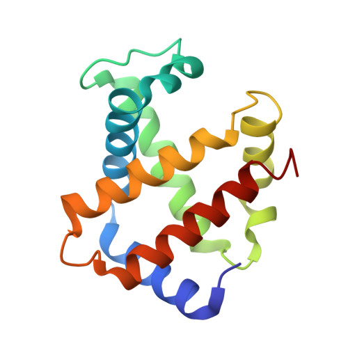
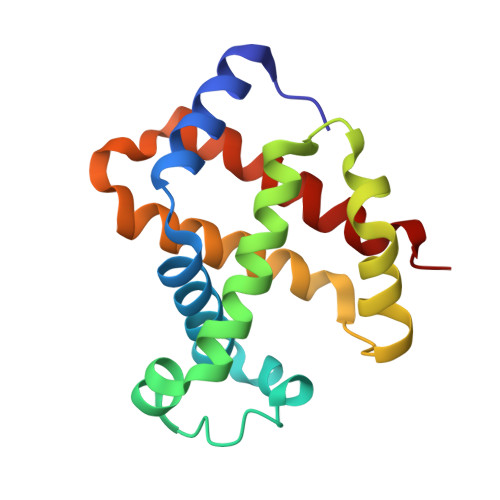
Crystal structure of haemoglobin from donkey (Equus asinus) at 3A resolution

Balasundaresan, D.Saraboji, K.Ponnuswamy, M.N.

(2006) Biochimie 88: 719-723

  • DOI: https://doi.org/10.1016/j.biochi.2006.01.001
  • Primary Citation of Related Structures:  
    1S0H

  • PubMed Abstract: 

    Haemoglobin from donkey was purified and crystallized in space group C2. The present donkey haemoglobin model comprises of two subunits alpha and beta. These alpha and beta subunits comprise of 141 and 146 amino acid residues, respectively, and the haem groups. The donkey haemoglobin differs from horse only in two amino acids of alpha-chain (His20 to Asn and Tyr24 to Phe) and these substitutions do not significantly change the secondary structural features of donkey haemoglobin. The haem group region and subunit contacts are closely resemble with that of horse methaemoglobin.


  • Organizational Affiliation

    Department of Crystallography and Biophysics, University of Madras, Guindy Campus, Chennai, 600025 India.


Macromolecules
Find similar proteins by:  (by identity cutoff)  |  3D Structure
Entity ID: 1
MoleculeChains Sequence LengthOrganismDetailsImage
Hemoglobin alpha chain141Equus asinusMutation(s): 0 
UniProt
Find proteins for P01959 (Equus asinus)
Explore P01959 
Go to UniProtKB:  P01959
Entity Groups  
Sequence Clusters30% Identity50% Identity70% Identity90% Identity95% Identity100% Identity
UniProt GroupP01959
Sequence Annotations
Expand
  • Reference Sequence
Find similar proteins by:  (by identity cutoff)  |  3D Structure
Entity ID: 2
MoleculeChains Sequence LengthOrganismDetailsImage
Hemoglobin beta chain146Equus asinusMutation(s): 0 
UniProt
Find proteins for D1MPT0 (Equus asinus)
Explore D1MPT0 
Go to UniProtKB:  D1MPT0
Entity Groups  
Sequence Clusters30% Identity50% Identity70% Identity90% Identity95% Identity100% Identity
UniProt GroupD1MPT0
Sequence Annotations
Expand
  • Reference Sequence
Small Molecules
Ligands 1 Unique
IDChains Name / Formula / InChI Key2D Diagram3D Interactions
HEM
Query on HEM

Download Ideal Coordinates CCD File 
C [auth A],
D [auth B]
PROTOPORPHYRIN IX CONTAINING FE
C34 H32 Fe N4 O4
KABFMIBPWCXCRK-RGGAHWMASA-L
Experimental Data & Validation

Experimental Data

  • Method: X-RAY DIFFRACTION
  • Resolution: 3.00 Å
  • R-Value Free: 0.285 
  • R-Value Work: 0.181 
  • R-Value Observed: 0.186 
  • Space Group: C 1 2 1
Unit Cell:
Length ( Å )Angle ( ˚ )
a = 107.664α = 90
b = 63.084β = 111.75
c = 54.042γ = 90
Software Package:
Software NamePurpose
REFMACrefinement
MAR345data collection
SCALEPACKdata scaling
AMoREphasing

Structure Validation

View Full Validation Report


Ligand Structure Quality Assessment 


Entry History 

Deposition Data

Revision History  (Full details and data files)

  • Version 1.0: 2005-02-01
    Type: Initial release
  • Version 1.1: 2008-04-29
    Changes: Version format compliance
  • Version 1.2: 2011-07-13
    Changes: Version format compliance
  • Version 1.3: 2017-10-11
    Changes: Refinement description
  • Version 1.4: 2023-10-25
    Changes: Data collection, Database references, Derived calculations, Refinement description