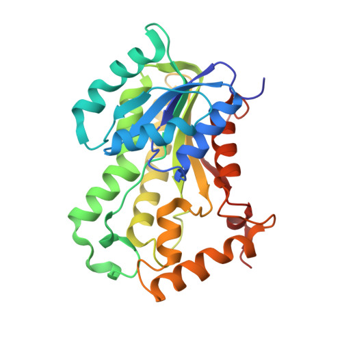
Targeting tuberculosis and malaria through inhibition of Enoyl reductase: compound activity and structural data.
Kuo, M.R., Morbidoni, H.R., Alland, D., Sneddon, S.F., Gourlie, B.B., Staveski, M.M., Leonard, M., Gregory, J.S., Janjigian, A.D., Yee, C., Musser, J.M., Kreiswirth, B., Iwamoto, H., Perozzo, R., Jacobs, W.R., Sacchettini, J.C., Fidock, D.A.(2003) J Biol Chem 278: 20851-20859
- PubMed: 12606558
- DOI: https://doi.org/10.1074/jbc.M211968200
- Primary Citation of Related Structures:
1P44, 1P45 - PubMed Abstract:
Tuberculosis and malaria together result in an estimated 5 million deaths annually. The spread of multidrug resistance in the most pathogenic causative agents, Mycobacterium tuberculosis and Plasmodium falciparum, underscores the need to identify active compounds with novel inhibitory properties. Although genetically unrelated, both organisms use a type II fatty-acid synthase system. Enoyl acyl carrier protein reductase (ENR), a key type II enzyme, has been repeatedly validated as an effective antimicrobial target. Using high throughput inhibitor screens with a combinatorial library, we have identified two novel classes of compounds with activity against the M. tuberculosis and P. falciparum enzyme (referred to as InhA and PfENR, respectively). The crystal structure of InhA complexed with NAD+ and one of the inhibitors was determined to elucidate the mode of binding. Structural analysis of InhA with the broad spectrum antimicrobial triclosan revealed a unique stoichiometry where the enzyme contained either a single triclosan molecule, in a configuration typical of other bacterial ENR:triclosan structures, or harbored two triclosan molecules bound to the active site. Significantly, these compounds do not require activation and are effective against wild-type and drug-resistant strains of M. tuberculosis and P. falciparum. Moreover, they provide broader chemical diversity and elucidate key elements of inhibitor binding to InhA for subsequent chemical optimization.
Organizational Affiliation:
Department of Biochemistry and Biophysics, Texas A & M University, College Station, Texas 77843, USA.


