1NNL

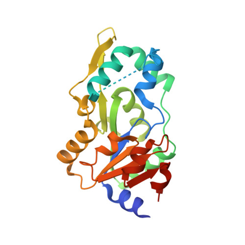
Crystal structure of Human Phosphoserine Phosphatase


Experimental Data Snapshot

  • Method: X-RAY DIFFRACTION
  • Resolution: 1.53 Å
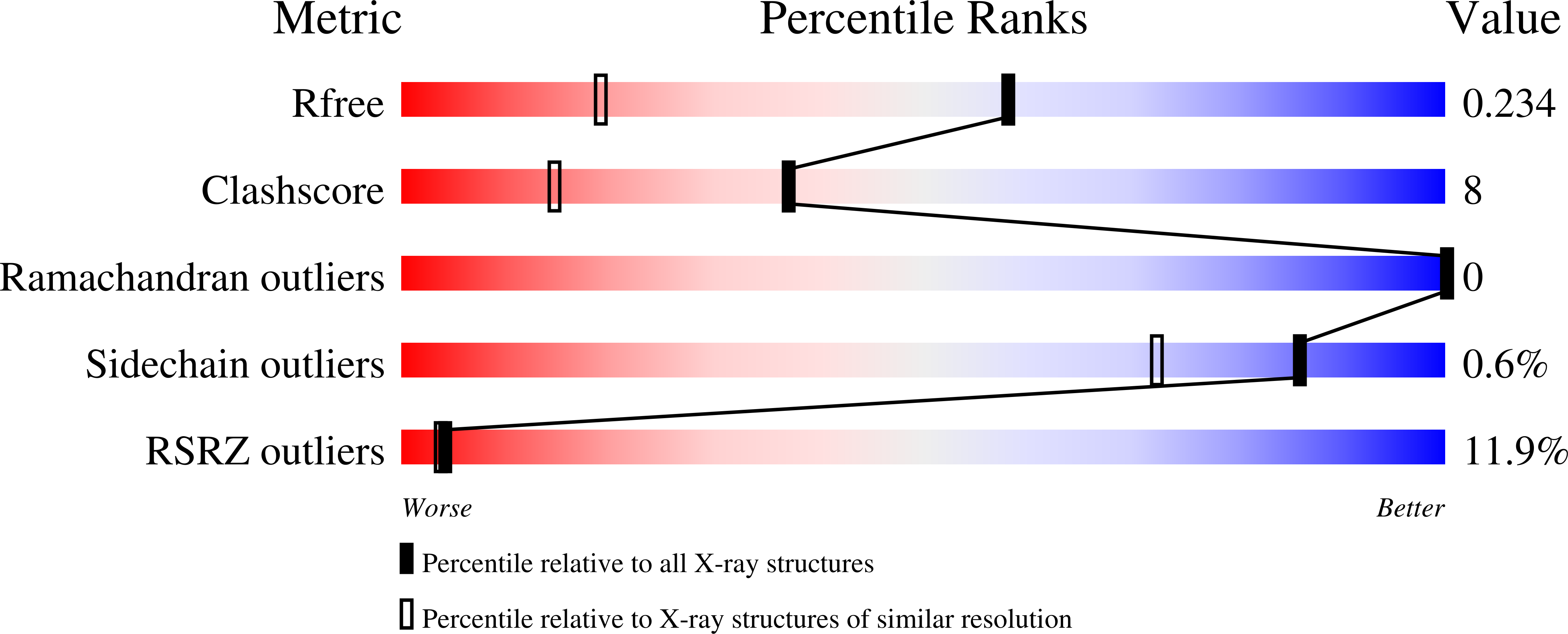
  • R-Value Free: 0.234 
  • R-Value Work: 0.217 
  • R-Value Observed: 0.217 

wwPDB Validation   3D Report Full Report


This is version 1.3 of the entry. See complete history


Literature

High-resolution structure of human phosphoserine phosphatase in open conformation.

Peeraer, Y.Rabijns, A.Verboven, C.Collet, J.F.Van Schaftingen, E.De Ranter, C.

(2003) Acta Crystallogr D Biol Crystallogr 59: 971-977

  • DOI: https://doi.org/10.1107/s0907444903005407
  • Primary Citation of Related Structures:  
    1NNL

  • PubMed Abstract: 

    The crystal structure of human phosphoserine phosphatase (HPSP) in the open conformation has been determined at a resolution of 1.53 A. The crystals are orthorhombic, belonging to space group C222(1), with unit-cell parameters a = 49.03, b = 130.25, c = 157.29 A. The asymmetric unit contains two molecules. Phase information was derived from a multiwavelength anomalous dispersion (MAD) experiment conducted at three wavelengths using a selenomethionine-derivative crystal of HPSP. The structure was refined using CNS to a final crystallographic R value of 21.6% (R(free) = 23.4%). HPSP is a dimeric enzyme responsible for the third and final step of the l-serine biosynthesis pathway. It catalyses the Mg2+-dependent hydrolysis of l-phosphoserine. Recently, the structure of HPSP in complex with an inhibitor bound to the active site has been reported to be the open conformation of the enzyme. Here, the structure of HPSP is reported in the absence of substrate in the active site. Evidence is presented that HPSP in an uncomplexed form is in an even more open conformation than in the inhibitor complex. In this state, the enzyme is partially unfolded to allow the substrate to enter the active site. Binding of the substrate causes HPSP to shift to the closed conformation by stabilizing the partially unfolded region. In the present structure a Ca2+ ion is bound to the active site and an explanation is given why HPSP is not active when in the active site Mg2+ is replaced by a Ca2+ ion.


  • Organizational Affiliation

    Laboratory for Analytical Chemistry and Medicinal Physicochemistry, Faculty of Pharmaceutical Sciences, K.U. Leuven, E. Van Evenstraat 4, B-3000 Leuven, Belgium.


Macromolecules
Find similar proteins by:  (by identity cutoff)  |  3D Structure
Entity ID: 1
MoleculeChains Sequence LengthOrganismDetailsImage
L-3-phosphoserine phosphatase
A, B
225Homo sapiensMutation(s): 0 
EC: 3.1.3.3
UniProt & NIH Common Fund Data Resources
Find proteins for P78330 (Homo sapiens)
Explore P78330 
Go to UniProtKB:  P78330
PHAROS:  P78330
GTEx:  ENSG00000146733 
Entity Groups  
Sequence Clusters30% Identity50% Identity70% Identity90% Identity95% Identity100% Identity
UniProt GroupP78330
Sequence Annotations
Expand
  • Reference Sequence
Small Molecules
Ligands 2 Unique
IDChains Name / Formula / InChI Key2D Diagram3D Interactions
CA
Query on CA

Download Ideal Coordinates CCD File 
F [auth A],
J [auth B],
K [auth B]
CALCIUM ION
Ca
BHPQYMZQTOCNFJ-UHFFFAOYSA-N
CL
Query on CL

Download Ideal Coordinates CCD File 
C [auth A]
D [auth A]
E [auth A]
G [auth B]
H [auth B]
C [auth A],
D [auth A],
E [auth A],
G [auth B],
H [auth B],
I [auth B]
CHLORIDE ION
Cl
VEXZGXHMUGYJMC-UHFFFAOYSA-M
Experimental Data & Validation

Experimental Data

  • Method: X-RAY DIFFRACTION
  • Resolution: 1.53 Å
  • R-Value Free: 0.234 
  • R-Value Work: 0.217 
  • R-Value Observed: 0.217 
  • Space Group: C 2 2 21
Unit Cell:
Length ( Å )Angle ( ˚ )
a = 49.033α = 90
b = 130.249β = 90
c = 157.293γ = 90
Software Package:
Software NamePurpose
CNSrefinement
DENZOdata reduction
SCALEPACKdata scaling
SHARPphasing

Structure Validation

View Full Validation Report


Entry History 

Deposition Data

Revision History  (Full details and data files)

  • Version 1.0: 2003-06-03
    Type: Initial release
  • Version 1.1: 2008-04-29
    Changes: Version format compliance
  • Version 1.2: 2011-07-13
    Changes: Version format compliance
  • Version 1.3: 2024-03-13
    Changes: Data collection, Database references, Derived calculations