1IER

CUBIC CRYSTAL STRUCTURE OF NATIVE HORSE SPLEEN FERRITIN


Experimental Data Snapshot

  • Method: X-RAY DIFFRACTION
  • Resolution: 2.26 Å
  • R-Value Work: 0.187 
  • R-Value Observed: 0.187 

Starting Model: experimental
View more details

wwPDB Validation   3D Report Full Report


This is version 1.5 of the entry. See complete history


Literature

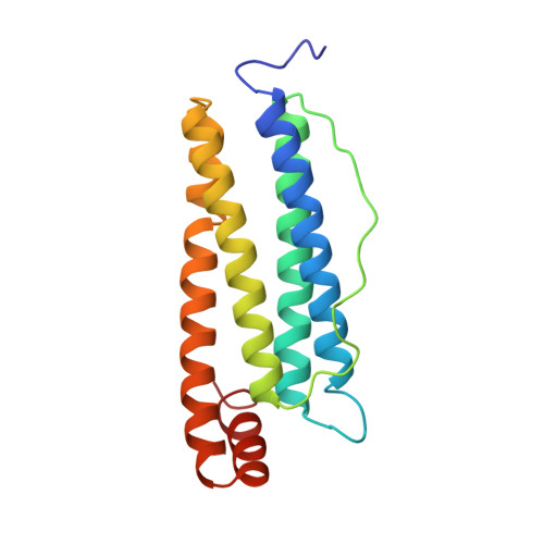
Comparison of the structures of the cubic and tetragonal forms of horse-spleen apoferritin.

Granier, T.Gallois, B.Dautant, A.Langlois d'Estaintot, B.Precigoux, G.

(1997) Acta Crystallogr D Biol Crystallogr 53: 580-587

  • DOI: https://doi.org/10.1107/S0907444997003314
  • Primary Citation of Related Structures:  
    1IER, 1IES

  • PubMed Abstract: 

    Horse-spleen apoferritin is known to crystallize in three different space groups, cubic F432, tetragonal P42(1)2 and orthorhombic P2(1)2(1)2. A structure comparison of the cubic and tetragonal forms is presented here. Both crystal forms were obtained by the vapor-diffusion technique and data were collected at 2.26 A (cubic crystal) and 2.60 A (tetragonal crystal) resolution. Two main differences were observed between these crystal structures: (i) whereas intermolecular contacts only involve salt-bridge type interactions via cadmium ions in the cubic structure, two types of interactions are observed in the tetragonal crystal (cadmium-ion-mediated salt bridges and hydrogen-bonding interactions) and (ii) cadmium ions bound in the threefold axes of ferritin molecules exhibit lower site-occupation factors in the tetragonal structure than in the cubic one.


  • Organizational Affiliation

    Unité de Biophysique Structurale, EP534 CNRS, Université Bordeaux 1, Talence, France.


Macromolecules
Find similar proteins by:  (by identity cutoff)  |  3D Structure
Entity ID: 1
MoleculeChains Sequence LengthOrganismDetailsImage
FERRITIN174Equus caballusMutation(s): 0 
UniProt
Find proteins for P02791 (Equus caballus)
Explore P02791 
Go to UniProtKB:  P02791
Entity Groups  
Sequence Clusters30% Identity50% Identity70% Identity90% Identity95% Identity100% Identity
UniProt GroupP02791
Sequence Annotations
Expand
  • Reference Sequence
Experimental Data & Validation

Experimental Data

  • Method: X-RAY DIFFRACTION
  • Resolution: 2.26 Å
  • R-Value Work: 0.187 
  • R-Value Observed: 0.187 
  • Space Group: F 4 3 2
Unit Cell:
Length ( Å )Angle ( ˚ )
a = 182.9α = 90
b = 182.9β = 90
c = 182.9γ = 90
Software Package:
Software NamePurpose
X-PLORmodel building
X-PLORrefinement
MOSFLMdata reduction
CCP4data scaling
ROTAVATAdata scaling
X-PLORphasing

Structure Validation

View Full Validation Report


Entry History 

Deposition Data

Revision History  (Full details and data files)

  • Version 1.0: 1997-01-11
    Type: Initial release
  • Version 1.1: 2008-03-05
    Changes: Version format compliance
  • Version 1.2: 2011-07-13
    Changes: Derived calculations, Version format compliance
  • Version 1.3: 2018-06-27
    Changes: Data collection, Derived calculations, Other, Refinement description
  • Version 1.4: 2023-08-09
    Changes: Database references, Derived calculations, Refinement description
  • Version 1.5: 2024-05-22
    Changes: Data collection