1H1H

Crystal Structure of Eosinophil Cationic Protein in Complex with 2',5'-ADP at 2.0 A resolution Reveals the Details of the Ribonucleolytic Active site


Experimental Data Snapshot

  • Method: X-RAY DIFFRACTION
  • Resolution: 2.00 Å
  • R-Value Free: 0.210 
  • R-Value Work: 0.205 
  • R-Value Observed: 0.205 

Starting Model: experimental
View more details

wwPDB Validation   3D Report Full Report


This is version 1.4 of the entry. See complete history


Literature

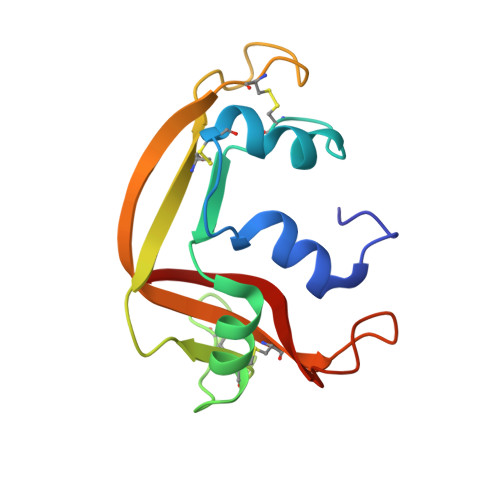
The Crystal Structure of Eosinophil Cationic Protein in Complex with 2'5'-Adp at 2.0 A Resolution Reveals the Details of the Ribonucleolytic Active Site

Mohan, C.G.Boix, E.Evans, H.R.Nikolovski, Z.Nogues, M.V.Cuchillo, C.M.Acharya, K.R.

(2002) Biochemistry 41: 12100

  • DOI: https://doi.org/10.1021/bi0264521
  • Primary Citation of Related Structures:  
    1H1H

  • PubMed Abstract: 

    Eosinophil cationic protein (ECP) is a component of the eosinophil granule matrix. It shows marked toxicity against helminth parasites, bacteria single-stranded RNA viruses, and host epithelial cells. Secretion of human ECP is related to eosinophil-associated allergic, asthmatic, and inflammatory diseases. ECP belongs to the pancreatic ribonuclease superfamily of proteins, and the crystal structure of ECP in the unliganded form (determined previously) exhibited a conserved RNase A fold [Boix, E., et al. (1999) Biochemistry 38, 16794-16801]. We have now determined a high-resolution (2.0 A) crystal structure of ECP in complex with adenosine 2',5'-diphosphate (2',5'-ADP) which has revealed the details of the ribonucleolytic active site. Residues Gln-14, His-15, and Lys-38 make hydrogen bond interactions with the phosphate at the P(1) site, while His-128 interacts with the purine ring at the B(2) site. A new phosphate binding site, P(-)(1), has been identified which involves Arg-34. This study is the first detailed structural analysis of the nucleotide recognition site in ECP and provides a starting point for the understanding of its substrate specificity and low catalytic efficiency compared with that of the eosinophil-derived neurotoxin (EDN), a close homologue.


  • Organizational Affiliation

    Department of Biology and Biochemistry, University of Bath, Claverton Down, Bath BA2 7AY, U.K.


Macromolecules
Find similar proteins by:  (by identity cutoff)  |  3D Structure
Entity ID: 1
MoleculeChains Sequence LengthOrganismDetailsImage
EOSINOPHIL CATIONIC PROTEIN134Homo sapiensMutation(s): 0 
EC: 3.1.27
Membrane Entity: Yes 
UniProt & NIH Common Fund Data Resources
Find proteins for P12724 (Homo sapiens)
Explore P12724 
Go to UniProtKB:  P12724
GTEx:  ENSG00000169397 
Entity Groups  
Sequence Clusters30% Identity50% Identity70% Identity90% Identity95% Identity100% Identity
UniProt GroupP12724
Sequence Annotations
Expand
  • Reference Sequence
Small Molecules
Ligands 1 Unique
IDChains Name / Formula / InChI Key2D Diagram3D Interactions
A2P
Query on A2P

Download Ideal Coordinates CCD File 
B [auth A]ADENOSINE-2'-5'-DIPHOSPHATE
C10 H15 N5 O10 P2
AEOBEOJCBAYXBA-KQYNXXCUSA-N
Binding Affinity Annotations 
IDSourceBinding Affinity
A2P PDBBind:  1H1H Ki: 6000 (nM) from 1 assay(s)
Experimental Data & Validation

Experimental Data

  • Method: X-RAY DIFFRACTION
  • Resolution: 2.00 Å
  • R-Value Free: 0.210 
  • R-Value Work: 0.205 
  • R-Value Observed: 0.205 
  • Space Group: P 63
Unit Cell:
Length ( Å )Angle ( ˚ )
a = 100.7α = 90
b = 100.7β = 90
c = 31.7γ = 120
Software Package:
Software NamePurpose
CNSrefinement
DENZOdata reduction
SCALEPACKdata scaling
AMoREphasing

Structure Validation

View Full Validation Report


Entry History 

Deposition Data

Revision History  (Full details and data files)

  • Version 1.0: 2002-10-03
    Type: Initial release
  • Version 1.1: 2011-05-08
    Changes: Version format compliance
  • Version 1.2: 2011-07-13
    Changes: Version format compliance
  • Version 1.3: 2019-10-16
    Changes: Data collection
  • Version 1.4: 2023-12-13
    Changes: Data collection, Database references, Refinement description