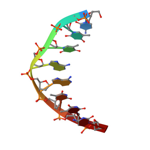
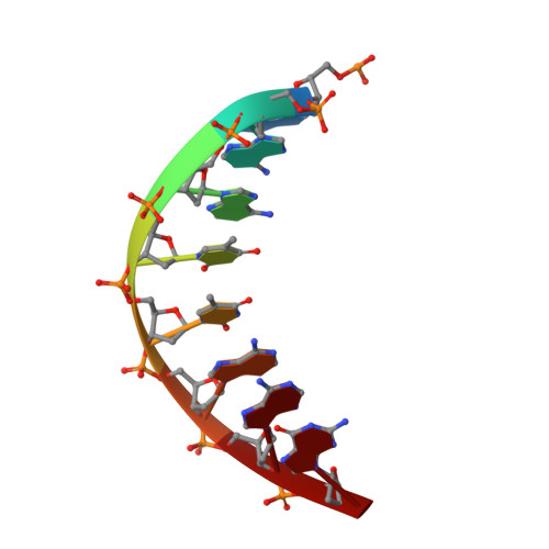
1ZTW

d(CTTAATTCGAATTAAG) complexed with Moloney Murine Leukemia Virus Reverse Transcriptase catalytic fragment


Experimental Data Snapshot

  • Method: X-RAY DIFFRACTION
  • Resolution: 1.80 Å
  • R-Value Free: 0.264 
  • R-Value Work: 0.227 
  • R-Value Observed: 0.227 

wwPDB Validation   3D Report Full Report


This is version 1.3 of the entry. See complete history


Literature

A host-guest approach for determining drug-DNA interactions: an example using netropsin.

Goodwin, K.D.Long, E.C.Georgiadis, M.M.

(2005) Nucleic Acids Res 33: 4106-4116

  • DOI: https://doi.org/10.1093/nar/gki717
  • Primary Citation of Related Structures:  
    1ZTT, 1ZTW

  • PubMed Abstract: 

    Netropsin is a well-characterized DNA minor groove binding compound that serves as a model for the study of drug-DNA interactions. Our laboratory has developed a novel host-guest approach to study drug-DNA interactions in which the host, the N-terminal fragment of Moloney murine leukemia virus reverse transcriptase (MMLV RT) is co-crystallized with a DNA oligonucleotide guest in the presence and absence of drug. We have co-crystallized netropsin with the RT fragment bound to the symmetric 16mer d(CTTAATTCGAATTAAG)2 and determined the structure of the complex at 1.85 A. In contrast to previously reported netropsin-DNA structures, our oligonucleotide contains two AATT sites that bind netropsin with flanking 5' and 3' sequences that are not symmetric. The asymmetric unit of the RT fragment-DNA-netropsin crystals contains one protein molecule and one-half of the 16mer with one netropsin molecule bound. The guanidinium moiety of netropsin binds in a narrow part of the minor groove, while the amidinium is bound in the widest region within the site. We compare this structure to other Class I netropsin-DNA structures and find that the asymmetry of minor groove widths in the AATT site contributes to the orientation of netropsin within the groove while hydrogen bonding patterns vary in the different structures.


  • Organizational Affiliation

    Department of Biochemistry and Molecular Biology, Indiana University School of Medicine, IN 46202, USA.


Macromolecules

Find similar proteins by:  (by identity cutoff)  |  3D Structure
Entity ID: 3
MoleculeChains Sequence LengthOrganismDetailsImage
Reverse transcriptaseC [auth A]255Moloney murine leukemia virusMutation(s): 0 
Gene Names: POL
EC: 2.7.7.49
UniProt
Find proteins for P03355 (Moloney murine leukemia virus (isolate Shinnick))
Explore P03355 
Go to UniProtKB:  P03355
Entity Groups  
Sequence Clusters30% Identity50% Identity70% Identity90% Identity95% Identity100% Identity
UniProt GroupP03355
Sequence Annotations
Expand
  • Reference Sequence

Find similar nucleic acids by:  Sequence   |   3D Structure  

Entity ID: 1
MoleculeChains LengthOrganismImage
CTTAATTCA [auth B]8N/A
Sequence Annotations
Expand
  • Reference Sequence

Find similar nucleic acids by:  Sequence   |   3D Structure  

Entity ID: 2
MoleculeChains LengthOrganismImage
GAATTAAGB [auth G]8N/A
Sequence Annotations
Expand
  • Reference Sequence
Experimental Data & Validation

Experimental Data

  • Method: X-RAY DIFFRACTION
  • Resolution: 1.80 Å
  • R-Value Free: 0.264 
  • R-Value Work: 0.227 
  • R-Value Observed: 0.227 
  • Space Group: P 21 21 2
Unit Cell:
Length ( Å )Angle ( ˚ )
a = 54.934α = 90
b = 145.746β = 90
c = 46.855γ = 90
Software Package:
Software NamePurpose
HKL-2000data collection
SCALEPACKdata scaling
AMoREphasing
CNSrefinement
HKL-2000data reduction

Structure Validation

View Full Validation Report


Entry History 

Deposition Data

Revision History  (Full details and data files)

  • Version 1.0: 2005-08-30
    Type: Initial release
  • Version 1.1: 2008-04-30
    Changes: Version format compliance
  • Version 1.2: 2011-07-13
    Changes: Version format compliance
  • Version 1.3: 2024-02-14
    Changes: Data collection, Database references