1ZBB

Structure of the 4_601_167 Tetranucleosome


Experimental Data Snapshot

  • Method: X-RAY DIFFRACTION
  • Resolution: 9.00 Å
  • R-Value Work: 0.386 
  • R-Value Observed: 0.386 

wwPDB Validation   3D Report Full Report


This is version 1.4 of the entry. See complete history


Literature

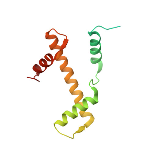
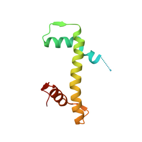
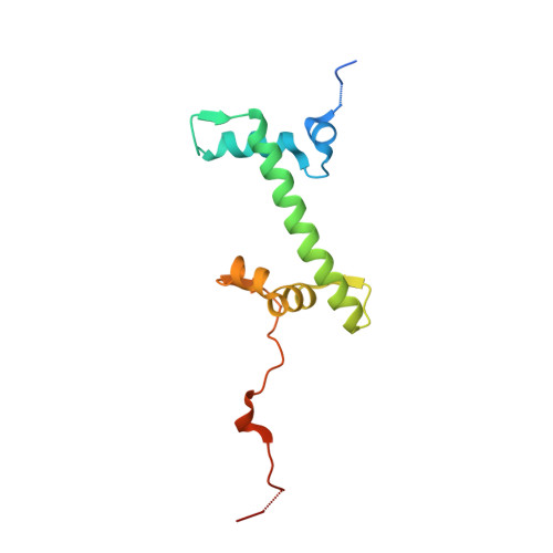
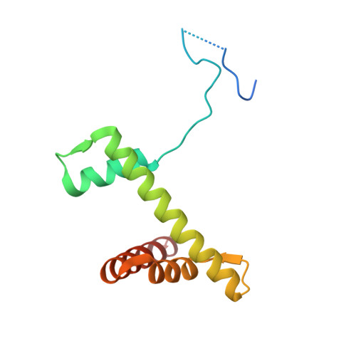
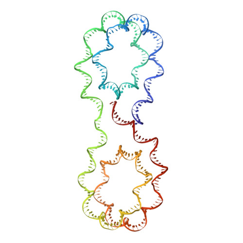
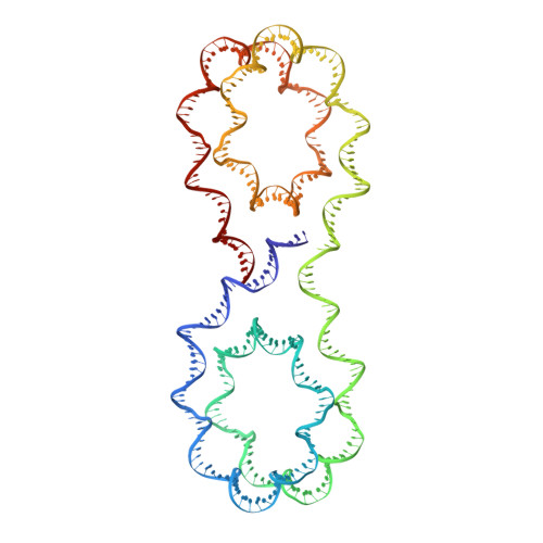
X-ray structure of a tetranucleosome and its implications for the chromatin fibre.

Schalch, T.Duda, S.Sargent, D.F.Richmond, T.J.

(2005) Nature 436: 138-141

  • DOI: https://doi.org/10.1038/nature03686
  • Primary Citation of Related Structures:  
    1ZBB

  • PubMed Abstract: 

    DNA in eukaryotic chromosomes is organized in arrays of nucleosomes compacted into chromatin fibres. This higher-order structure of nucleosomes is the substrate for DNA replication, recombination, transcription and repair. Although the structure of the nucleosome core is known at near-atomic resolution, even the most fundamental information about the organization of nucleosomes in the fibre is controversial. Here we report the crystal structure of an oligonucleosome (a compact tetranucleosome) at 9 A resolution, solved by molecular replacement using the nucleosome core structure. The structure shows that linker DNA zigzags back and forth between two stacks of nucleosome cores, which form a truncated two-start helix, and does not follow a path compatible with a one-start solenoidal helix. The length of linker DNA is most probably buffered by stretching of the DNA contained in the nucleosome cores. We have built continuous fibre models by successively stacking tetranucleosomes one on another. The resulting models are nearly fully compacted and most closely resemble the previously described crossed-linker model. They suggest that the interfaces between nucleosomes along a single helix start are polymorphic.


  • Organizational Affiliation

    ETH Zürich, Institute for Molecular Biology and Biophysics, ETH-Hönggerberg, CH-8093 Zürich, Switzerland.


Macromolecules

Find similar proteins by:  (by identity cutoff)  |  3D Structure
Entity ID: 3
MoleculeChains Sequence LengthOrganismDetailsImage
HISTONE H3C [auth A],
G [auth E],
K [auth a],
O [auth e]
135Xenopus laevisMutation(s): 0 
UniProt
Find proteins for P84233 (Xenopus laevis)
Explore P84233 
Go to UniProtKB:  P84233
Entity Groups  
Sequence Clusters30% Identity50% Identity70% Identity90% Identity95% Identity100% Identity
UniProt GroupP84233
Sequence Annotations
Expand
  • Reference Sequence
Find similar proteins by:  (by identity cutoff)  |  3D Structure
Entity ID: 4
MoleculeChains Sequence LengthOrganismDetailsImage
Histone H4D [auth B],
H [auth F],
L [auth b],
P [auth f]
102Xenopus laevisMutation(s): 0 
UniProt
Find proteins for P62799 (Xenopus laevis)
Explore P62799 
Go to UniProtKB:  P62799
Entity Groups  
Sequence Clusters30% Identity50% Identity70% Identity90% Identity95% Identity100% Identity
UniProt GroupP62799
Sequence Annotations
Expand
  • Reference Sequence
Find similar proteins by:  (by identity cutoff)  |  3D Structure
Entity ID: 5
MoleculeChains Sequence LengthOrganismDetailsImage
Histone H2A.1E [auth C],
I [auth G],
M [auth c],
Q [auth g]
129Xenopus laevisMutation(s): 0 
UniProt
Find proteins for P06897 (Xenopus laevis)
Explore P06897 
Go to UniProtKB:  P06897
Entity Groups  
Sequence Clusters30% Identity50% Identity70% Identity90% Identity95% Identity100% Identity
UniProt GroupP06897
Sequence Annotations
Expand
  • Reference Sequence
Find similar proteins by:  (by identity cutoff)  |  3D Structure
Entity ID: 6
MoleculeChains Sequence LengthOrganismDetailsImage
Histone H2B.1F [auth D],
J [auth H],
N [auth d],
R [auth h]
125Xenopus laevisMutation(s): 0 
UniProt
Find proteins for P02281 (Xenopus laevis)
Explore P02281 
Go to UniProtKB:  P02281
Entity Groups  
Sequence Clusters30% Identity50% Identity70% Identity90% Identity95% Identity100% Identity
UniProt GroupP02281
Sequence Annotations
Expand
  • Reference Sequence
Find similar nucleic acids by:  (by identity cutoff)  |  3D Structure
Entity ID: 1
MoleculeChains LengthOrganismImage
DNA STRAND 1 (ARBITRARY MODEL SEQUENCE)A [auth I]347N/A
Sequence Annotations
Expand
  • Reference Sequence
Find similar nucleic acids by:  (by identity cutoff)  |  3D Structure
Entity ID: 2
MoleculeChains LengthOrganismImage
DNA STRAND 2 (ARBITRARY MODEL SEQUENCE)B [auth J]347N/A
Sequence Annotations
Expand
  • Reference Sequence
Experimental Data & Validation

Experimental Data

  • Method: X-RAY DIFFRACTION
  • Resolution: 9.00 Å
  • R-Value Work: 0.386 
  • R-Value Observed: 0.386 
  • Space Group: I 2 2 2
Unit Cell:
Length ( Å )Angle ( ˚ )
a = 127.675α = 90
b = 168.445β = 90
c = 237.126γ = 90
Software Package:
Software NamePurpose
BEASTmodel building
REFMACrefinement
BEASTphasing

Structure Validation

View Full Validation Report


Entry History 

Deposition Data

Revision History  (Full details and data files)

  • Version 1.0: 2005-07-12
    Type: Initial release
  • Version 1.1: 2008-04-30
    Changes: Version format compliance
  • Version 1.2: 2011-07-13
    Changes: Version format compliance
  • Version 1.3: 2014-05-14
    Changes: Other
  • Version 1.4: 2023-10-25
    Changes: Data collection, Database references, Refinement description