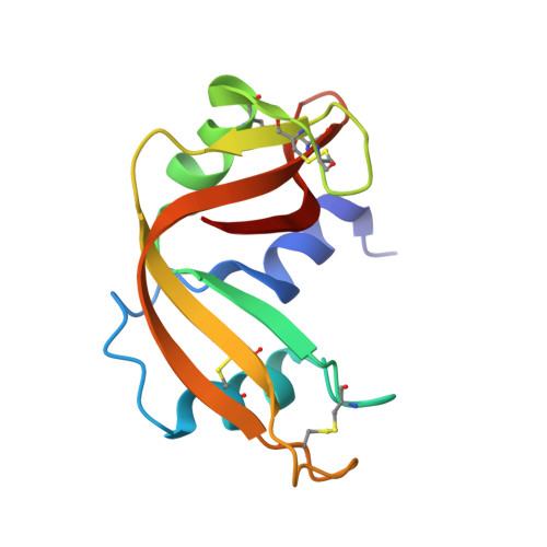
The binding of IMP to Ribonuclease A
Hatzopoulos, G.N., Leonidas, D.D., Kardakaris, R., Kobe, J., Oikonomakos, N.G.(2005) FEBS J 272: 3988-4001
- PubMed: 16045769
- DOI: https://doi.org/10.1111/j.1742-4658.2005.04822.x
- Primary Citation of Related Structures:
1Z6D, 1Z6S - PubMed Abstract:
The binding of inosine 5' phosphate (IMP) to ribonuclease A has been studied by kinetic and X-ray crystallographic experiments at high (1.5 A) resolution. IMP is a competitive inhibitor of the enzyme with respect to C>p and binds to the catalytic cleft by anchoring three IMP molecules in a novel binding mode. The three IMP molecules are connected to each other by hydrogen bond and van der Waals interactions and collectively occupy the B1R1P1B2P0P(-1) region of the ribonucleolytic active site. One of the IMP molecules binds with its nucleobase in the outskirts of the B2 subsite and interacts with Glu111 while its phosphoryl group binds in P1. Another IMP molecule binds by following the retro-binding mode previously observed only for guanosines with its nucleobase at B1 and the phosphoryl group in P(-1). The third IMP molecule binds in a novel mode towards the C-terminus. The RNase A-IMP complex provides structural evidence for the functional components of subsite P(-1) while it further supports the role inferred by other studies to Asn71 as the primary structural determinant for the adenine specificity of the B2 subsite. Comparative structural analysis of the IMP and AMP complexes highlights key aspects of the specificity of the base binding subsites of RNase A and provides a structural explanation for their potencies. The binding of IMP suggests ways to develop more potent inhibitors of the pancreatic RNase superfamily using this nucleotide as the starting point.
Organizational Affiliation:
Institute of Organic & Pharmaceutical Chemistry, The National Hellenic Research Foundation, Athens, Greece.