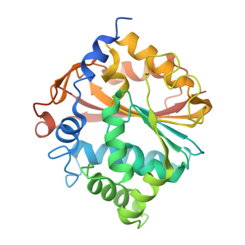
1YZ3

Structure of human pnmt complexed with cofactor product adohcy and inhibitor SK&F 64139


Experimental Data Snapshot

  • Method: X-RAY DIFFRACTION
  • Resolution: 2.40 Å
  • R-Value Free: 0.243 
  • R-Value Work: 0.214 
  • R-Value Observed: 0.214 

Starting Model: experimental
View more details

wwPDB Validation   3D Report Full Report


Ligand Structure Quality Assessment 


This is version 1.4 of the entry. See complete history


Literature

Structural, mutagenic, and kinetic analysis of the binding of substrates and inhibitors of human phenylethanolamine N-methyltransferase

Wu, Q.Gee, C.L.Lin, F.Tyndall, J.D.Martin, J.L.Grunewald, G.L.McLeish, M.J.

(2005) J Med Chem 48: 7243-7252

  • DOI: https://doi.org/10.1021/jm050568o
  • Primary Citation of Related Structures:  
    1YZ3

  • PubMed Abstract: 

    The X-ray structure of human phenylethanolamine N-methyltransferase (hPNMT) complexed with its product, S-adenosyl-L-homocysteine (4), and the most potent inhibitor reported to date, SK&F 64139 (7), was used to identify the residues involved in inhibitor binding. Four of these residues, Val53, Lys57, Glu219 and Asp267, were replaced, in turn, with alanine. All variants had increased Km values for phenylethanolamine (10), but only D267A showed a noteworthy (20-fold) decrease in its kcat value. Both WT hPNMT and D267A had similar kcat values for a rigid analogue, anti-9-amino-6-(trifluoromethyl)benzonorbornene (12), suggesting that Asp267 plays an important role in positioning the substrate but does not participate directly in catalysis. The Ki values for the binding of inhibitors such as 7 to the E219A and D267A variants increased by 2-3 orders of magnitude. Further, the inhibitors were shown to bind up to 50-fold more tightly in the presence of S-adenosyl-L-methionine (3), suggesting that the binding of the latter brings about a conformational change in the enzyme.


  • Organizational Affiliation

    Department of Medicinal Chemistry, University of Michigan, 428 Church Street, Ann Arbor, Michigan 48109, USA.


Macromolecules
Find similar proteins by:  (by identity cutoff)  |  3D Structure
Entity ID: 1
MoleculeChains Sequence LengthOrganismDetailsImage
Phenylethanolamine N-methyltransferase
A, B
289Homo sapiensMutation(s): 0 
EC: 2.1.1.28
UniProt & NIH Common Fund Data Resources
Find proteins for P11086 (Homo sapiens)
Explore P11086 
Go to UniProtKB:  P11086
PHAROS:  P11086
GTEx:  ENSG00000141744 
Entity Groups  
Sequence Clusters30% Identity50% Identity70% Identity90% Identity95% Identity100% Identity
UniProt GroupP11086
Sequence Annotations
Expand
  • Reference Sequence
Small Molecules
Binding Affinity Annotations 
IDSourceBinding Affinity
SAH BindingDB:  1YZ3 Ki: 1.40e+4 (nM) from 1 assay(s)
SKA PDBBind:  1YZ3 Ki: 1.55 (nM) from 1 assay(s)
BindingDB:  1YZ3 Ki: min: 1.55, max: 300 (nM) from 4 assay(s)
IC50: min: 3, max: 1.00e+4 (nM) from 3 assay(s)
Experimental Data & Validation

Experimental Data

  • Method: X-RAY DIFFRACTION
  • Resolution: 2.40 Å
  • R-Value Free: 0.243 
  • R-Value Work: 0.214 
  • R-Value Observed: 0.214 
  • Space Group: P 43 21 2
Unit Cell:
Length ( Å )Angle ( ˚ )
a = 93.9α = 90
b = 93.9β = 90
c = 188.3γ = 90
Software Package:
Software NamePurpose
d*TREKdata reduction
CNSrefinement
d*TREKdata scaling
CNSphasing

Structure Validation

View Full Validation Report


Ligand Structure Quality Assessment 


Entry History 

Deposition Data

Revision History  (Full details and data files)

  • Version 1.0: 2006-02-07
    Type: Initial release
  • Version 1.1: 2008-04-30
    Changes: Version format compliance
  • Version 1.2: 2011-07-13
    Changes: Version format compliance
  • Version 1.3: 2017-10-11
    Changes: Refinement description
  • Version 1.4: 2023-10-25
    Changes: Data collection, Database references, Derived calculations, Refinement description