High-resolution refinement of yeast iso-1-cytochrome c and comparisons with other eukaryotic cytochromes c.
Louie, G.V., Brayer, G.D.(1990) J Mol Biol 214: 527-555
- PubMed: 2166169
- DOI: https://doi.org/10.1016/0022-2836(90)90197-T
- Primary Citation of Related Structures:
1YCC - PubMed Abstract:
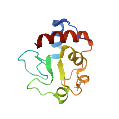
The structure of yeast iso-1-cytochrome c has been refined against X-ray diffraction data to a nominal resolution of 1.23 A. The atomic model contains 893 protein atoms, as well as 116 water molecules and one sulfate anion. Also included in the refinement are 886 hydrogen atoms belonging to the protein molecule. The crystallographic R-factor is 0.192 for the 12,513 reflections with F greater than or equal to 3 sigma (F) in the resolution range 6.0 to 1.23 A. Co-ordinate accuracy is estimated to be better than 0.18 A. The iso-1-cytochrome c molecule has the typical cytochrome c fold, with the polypeptide chain organized into a series of alpha-helices and reverse turns that serve to envelop the heme prosthetic group in a hydrophobic pocket. Inspection of the conformations of helices in the molecule shows that the local environments of the helices, in particular the presence of intrahelical threonine residues, cause distortions from ideal alpha-helical geometry. Analysis of the internal mobility of iso-1-cytochrome c, based on refined crystallographic temperature factors, shows that the most rigid parts of the molecule are those that are closely associated with the heme group. The degree of saturation of hydrogen-bonding potential is high, with 90% of all polar atoms found to participate in hydrogen bonding. The geometry of intramolecular hydrogen bonds is typical of that observed in other high-resolution protein structures. The 116 water molecules present in the model represent about 41% of those expected to be present in the asymmetric unit. The majority of the water molecules are organized into a small number of hydrogen-bonding networks that are anchored to the protein surface. Comparison of the structure of yeast iso-1-cytochrome c with those of tuna and rice cytochromes c shows that these three molecules have very high structural similarity, with the atomic packing in the heme crevice region being particularly highly conserved. Large conformational differences that are observed between these cytochromes c can be explained by amino acid substitutions. Additional subtle differences in the positioning of the side-chains of several highly conserved residues are also observed and occur due to unique features in the local environments of each cytochrome c molecule.(ABSTRACT TRUNCATED AT 400 WORDS)
Organizational Affiliation:
Department of Biochemistry, University of British Columbia, Vancouver, Canada.