1XDG

X-ray structure of LFA-1 I-domain in complex with LFA878 at 2.1A resolution


Experimental Data Snapshot

  • Method: X-RAY DIFFRACTION
  • Resolution: 2.10 Å
  • R-Value Free: 0.213 
  • R-Value Work: 0.191 
  • R-Value Observed: 0.192 

wwPDB Validation   3D Report Full Report


Ligand Structure Quality Assessment 


This is version 1.4 of the entry. See complete history


Literature

Improved lymphocyte function-associated antigen-1 (LFA-1) inhibition by statin derivatives: molecular basis determined by x-ray analysis and monitoring of LFA-1 conformational changes in vitro and ex vivo

Weitz-Schmidt, G.Welzenbach, K.Dawson, J.Kallen, J.

(2004) J Biol Chem 279: 46764-46771

  • DOI: https://doi.org/10.1074/jbc.M407951200
  • Primary Citation of Related Structures:  
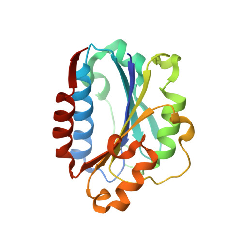
    1XDD, 1XDG

  • PubMed Abstract: 

    The integrin lymphocyte function-associated antigen-1 (LFA-1) (alphaLbeta2; CD11a/CD18) plays an important role in leukocyte migration and T cell activation. LFA-1 is inhibited by the cholesterol-lowering drug lovastatin, which binds to an allosteric site of the alphaL I domain termed the lovastatin site (L-site). Here we report for the first time the x-ray structures of the LFA-1 I domain complexed with derivatives of lovastatin optimized for LFA-1 inhibition. This analysis identified two new subpockets within the L-site occupied by chemical groups of the statin derivatives but not by lovastatin itself. Occupancy of these L-site subpockets led to distinct conformational changes in LFA-1, which were detectable by an epitope-monitoring assay. We utilized this assay to demonstrate improved LFA-1 inhibition in human blood in vitro and in blood samples from treated animals ex vivo. Moreover, we demonstrate that the novel lovastatin-derived LFA-1 inhibitor LFA878 exhibits potent anti-inflammatory effects in carrageenan-induced rat paw edema. In summary, the findings reported here extend the understanding of LFA-1 inhibition at the molecular level, allow for the identification and design of LFA-1 inhibitors of further enhanced potency, and support the expectation that LFA-1 inhibitors binding to the L-site will be of therapeutic value in treating inflammatory diseases.


  • Organizational Affiliation

    Novartis Institutes for BioMedical Research, CH-4002 Basel, Switzerland.


Macromolecules
Find similar proteins by:  (by identity cutoff)  |  3D Structure
Entity ID: 1
MoleculeChains Sequence LengthOrganismDetailsImage
Integrin alpha-L
A, B
188Homo sapiensMutation(s): 0 
UniProt & NIH Common Fund Data Resources
Find proteins for P20701 (Homo sapiens)
Explore P20701 
Go to UniProtKB:  P20701
PHAROS:  P20701
GTEx:  ENSG00000005844 
Entity Groups  
Sequence Clusters30% Identity50% Identity70% Identity90% Identity95% Identity100% Identity
UniProt GroupP20701
Sequence Annotations
Expand
  • Reference Sequence
Small Molecules
Ligands 2 Unique
IDChains Name / Formula / InChI Key2D Diagram3D Interactions
AB8
Query on AB8

Download Ideal Coordinates CCD File 
D [auth A],
F [auth B]
(1S,3R,8AS)-8-(2-{(4S,6S)-3-(4-HYDROXY-3-METHOXYBENZYL)-4-[2-(METHYLAMINO)-2-OXOETHYL]-2-OXO-1,3-OXAZINAN-6-YL}ETHYL)-3 ,7-DIMETHYL-1,2,3,7,8,8A-HEXAHYDRONAPHTHALEN-1-YL (2R)-2-METHYLBUTANOATE
C34 H48 N2 O7
XGCZRGJUCNQVCO-XFAJEYMZSA-N
MG
Query on MG

Download Ideal Coordinates CCD File 
C [auth A],
E [auth B]
MAGNESIUM ION
Mg
JLVVSXFLKOJNIY-UHFFFAOYSA-N
Binding Affinity Annotations 
IDSourceBinding Affinity
AB8 Binding MOAD:  1XDG IC50: 50 (nM) from 1 assay(s)
PDBBind:  1XDG IC50: 50 (nM) from 1 assay(s)
BindingDB:  1XDG IC50: min: 50, max: 250 (nM) from 2 assay(s)
Experimental Data & Validation

Experimental Data

  • Method: X-RAY DIFFRACTION
  • Resolution: 2.10 Å
  • R-Value Free: 0.213 
  • R-Value Work: 0.191 
  • R-Value Observed: 0.192 
  • Space Group: P 21 21 21
Unit Cell:
Length ( Å )Angle ( ˚ )
a = 71.9α = 90
b = 77.4β = 90
c = 92.3γ = 90
Software Package:
Software NamePurpose
REFMACrefinement
DENZOdata reduction
SCALEPACKdata scaling
X-PLORphasing

Structure Validation

View Full Validation Report


Ligand Structure Quality Assessment 


Entry History 

Deposition Data

Revision History  (Full details and data files)

  • Version 1.0: 2004-09-21
    Type: Initial release
  • Version 1.1: 2008-04-30
    Changes: Version format compliance
  • Version 1.2: 2011-07-13
    Changes: Version format compliance
  • Version 1.3: 2013-02-20
    Changes: Database references
  • Version 1.4: 2023-10-25
    Changes: Data collection, Database references, Derived calculations, Refinement description, Structure summary