1VLZ

UNCOUPLED PHOSPHORYLATION AND ACTIVATION IN BACTERIAL CHEMOTAXIS: THE 2.1 ANGSTROM STRUCTURE OF A THREONINE TO ISOLEUCINE MUTANT AT POSITION 87 OF CHEY


Experimental Data Snapshot

  • Method: X-RAY DIFFRACTION
  • Resolution: 2.05 Å
  • R-Value Observed: 0.156 

wwPDB Validation   3D Report Full Report


This is version 1.4 of the entry. See complete history


Literature

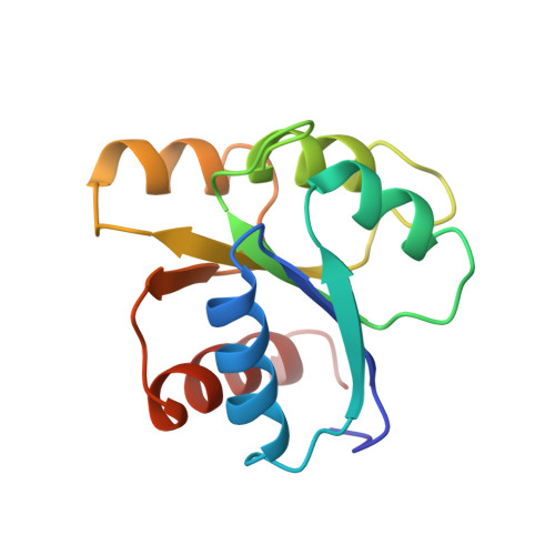
Uncoupled phosphorylation and activation in bacterial chemotaxis. The 2.1-A structure of a threonine to isoleucine mutant at position 87 of CheY.

Ganguli, S.Wang, H.Matsumura, P.Volz, K.

(1995) J Biol Chem 270: 17386-17393

  • Primary Citation of Related Structures:  
    1VLZ

  • PubMed Abstract: 

    Position 87 of the chemotaxis regulatory protein CheY is a highly conserved threonine/serine residue in the response regulator superfamily. A threonine 87 to isoleucine mutant in CheY, identified by its in vivo non-chemotactic phenotype, was also found to be phosphorylatable in vitro. These properties indicate that this mutant does not undergo activation upon phosphorylation. The x-ray crystallographic structure of the threonine to isoleucine CheY mutant has been solved and refined at 2.1-A resolution, to an R factor of 15.6%. Comparison with the wild-type, Mg(2+)-free CheY structure shows that the active site structure is retained, but there are significant localized differences in the backbone conformation distal from the substitution. The presence of the isoleucine side chain also restricts the rotational conformation of another conserved residue in the molecule, tyrosine at position 106. These results provide further evidence for a signaling surface remote from the phosphorylation site of the CheY molecule and implicate threonine 87 and other residues in the post-phosphorylation signaling events.


  • Organizational Affiliation

    Department of Microbiology and Immunology, University of Illinois, Chicago 60612, USA.


Macromolecules
Find similar proteins by:  (by identity cutoff)  |  3D Structure
Entity ID: 1
MoleculeChains Sequence LengthOrganismDetailsImage
CHEY
A, B
128Escherichia coliMutation(s): 0 
UniProt
Find proteins for P0AE67 (Escherichia coli (strain K12))
Explore P0AE67 
Go to UniProtKB:  P0AE67
Entity Groups  
Sequence Clusters30% Identity50% Identity70% Identity90% Identity95% Identity100% Identity
UniProt GroupP0AE67
Sequence Annotations
Expand
  • Reference Sequence
Experimental Data & Validation

Experimental Data

  • Method: X-RAY DIFFRACTION
  • Resolution: 2.05 Å
  • R-Value Observed: 0.156 
  • Space Group: P 1 21 1
Unit Cell:
Length ( Å )Angle ( ˚ )
a = 54.04α = 90
b = 72.47β = 109.06
c = 36.15γ = 90
Software Package:
Software NamePurpose
PROFFTrefinement
XENGENdata reduction

Structure Validation

View Full Validation Report


Entry History 

Deposition Data

Revision History  (Full details and data files)

  • Version 1.0: 1995-07-10
    Type: Initial release
  • Version 1.1: 2008-03-03
    Changes: Version format compliance
  • Version 1.2: 2011-07-13
    Changes: Version format compliance
  • Version 1.3: 2017-11-29
    Changes: Derived calculations, Other
  • Version 1.4: 2024-02-14
    Changes: Data collection, Database references