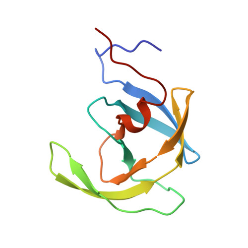
Structure of HOE/BAY 793 complexed to human immunodeficiency virus (HIV-1) protease in two different crystal forms--structure/function relationship and influence of crystal packing.
Lange-Savage, G., Berchtold, H., Liesum, A., Budt, K.H., Peyman, A., Knolle, J., Sedlacek, J., Fabry, M., Hilgenfeld, R.(1997) Eur J Biochem 248: 313-322
- PubMed: 9346283
- DOI: https://doi.org/10.1111/j.1432-1033.1997.00313.x
- Primary Citation of Related Structures:
1VIJ, 1VIK - PubMed Abstract:
Human immunodeficiency virus 1 (HIV-1) protease is a prime target in the search for drugs to combat the AIDS virus. The enzyme functions as a C2-symmetric dimer, cleaving the gag and gag-pol viral polyproteins at distinct sites. The possession of a twofold axis passing through the active site, has led to the design of C2-symmetrical inhibitors in the form of substrate-based transition-state analogs. One of the most active compounds of this class of inhibitors is HOE/BAY 793, which contains a vicinal diol central unit [Budt, K.-H., Hansen, J., Knolle, J., Meichsner, C., Paessens, A., Ruppert, D. & Stowasser, B. & Winkler, I. (1990) European Patent application EP0428,849; Budt, K.-H., Hansen, J., Knolle, J., Meichsner, C., Ruppert, D., Paessens, A. & Stowasser B. (1993) IXth International Conference on AIDS; Budt, K.-H., Peyman, A., Hansen, J., Knolle, J., Meichsner, C., Paessens, A., Ruppert, D. & Stowasser, B. (1995) Bioorg. Med. Chem. 3, 559-571.] The structure of this inhibitor bound to HIV-1 protease, in two different crystal forms, has been solved at 0.24-nm resolution using X-ray crystallography. In both forms, the details of the inhibitor-protease interactions revealed an overall asymmetric binding mode, especially between the central diol unit and the active-site aspartates. The main binding interactions comprise several specific H-bonds and hydrophobic contacts, which rationalize many of the characteristics of the structure/activity relationship in the class of vicinal diol inhibitors. In a general analysis of the mobility of the flap regions, which cover the active site and participate directly in binding, using our structures and the HIV protease models present in the Brookhaven databank, we found that in most structures the flexibility of the flaps is limited by local crystal contacts. However, in one of the structures presented here, no significant crystal contacts to the flap regions were present, and as a result the flexibility of the inhibitor bound flaps increased significantly. This suggests that the mobility and conformational flexibility of the flap residues are important in the functioning of HIV-1 protease, and must be considered in the future design of drugs against HIV protease and in structure-based drug design in general.
Organizational Affiliation:
Hoechst AG, Central Pharma Research, Frankfurt, Germany.


