1VFP

Crystal structure of the SR CA2+-ATPase with bound AMPPCP


Experimental Data Snapshot

  • Method: X-RAY DIFFRACTION
  • Resolution: 2.90 Å
  • R-Value Free: 0.287 
  • R-Value Work: 0.247 
  • R-Value Observed: 0.255 

wwPDB Validation   3D Report Full Report


This is version 1.3 of the entry. See complete history


Literature

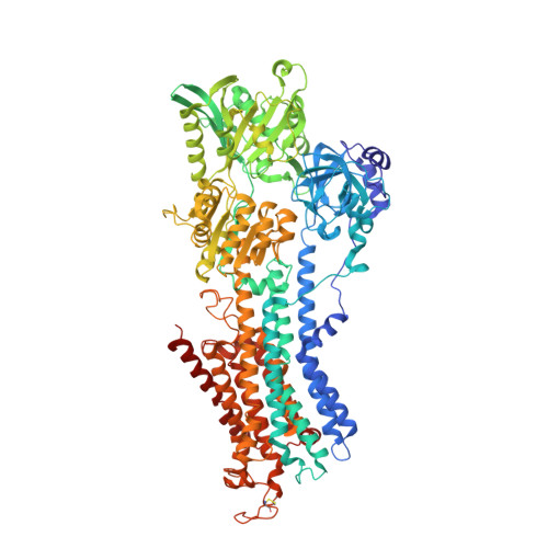
Crystal structure of the calcium pump with a bound ATP analogue.

Toyoshima, C.Mizutani, T.

(2004) Nature 430: 529-535

  • DOI: https://doi.org/10.1038/nature02680
  • Primary Citation of Related Structures:  
    1VFP

  • PubMed Abstract: 

    P-type ATPases are ATP-powered ion pumps that establish ion concentration gradients across cell and organelle membranes. Here, we describe the crystal structure of the Ca2+ pump of skeletal muscle sarcoplasmic reticulum, a representative member of the P-type ATPase superfamily, with an ATP analogue, a Mg2+ and two Ca2+ ions in the respective binding sites. In this state, the ATP analogue reorganizes the three cytoplasmic domains (A, N and P), which are widely separated without nucleotide, by directly bridging the N and P domains. The structure of the P-domain itself is altered by the binding of the ATP analogue and Mg2+. As a result, the A-domain is tilted so that one of the transmembrane helices moves to lock the cytoplasmic gate of the transmembrane Ca2+-binding sites. This appears to be the mechanism for occluding the bound Ca2+ ions, before releasing them into the lumen of the sarcoplasmic reticulum.


  • Organizational Affiliation

    Institute of Molecular and Cellular Biosciences, The University of Tokyo, Bunkyo-ku, Tokyo 113-0032, Japan. ct@iam.u-tokyo.ac.jp


Macromolecules
Find similar proteins by:  (by identity cutoff)  |  3D Structure
Entity ID: 1
MoleculeChains Sequence LengthOrganismDetailsImage
Sarcoplasmic/endoplasmic reticulum calcium ATPase 1
A, B
994Oryctolagus cuniculusMutation(s): 0 
EC: 3.6.3.8
Membrane Entity: Yes 
UniProt
Find proteins for P04191 (Oryctolagus cuniculus)
Explore P04191 
Go to UniProtKB:  P04191
Entity Groups  
Sequence Clusters30% Identity50% Identity70% Identity90% Identity95% Identity100% Identity
UniProt GroupP04191
Sequence Annotations
Expand
  • Reference Sequence
Experimental Data & Validation

Experimental Data

  • Method: X-RAY DIFFRACTION
  • Resolution: 2.90 Å
  • R-Value Free: 0.287 
  • R-Value Work: 0.247 
  • R-Value Observed: 0.255 
  • Space Group: P 1 21 1
Unit Cell:
Length ( Å )Angle ( ˚ )
a = 90.92α = 90
b = 123.62β = 107.21
c = 151.82γ = 90
Software Package:
Software NamePurpose
CNSrefinement
DENZOdata reduction
SCALEPACKdata scaling
CNSphasing

Structure Validation

View Full Validation Report


Entry History 

Deposition Data

Revision History  (Full details and data files)

  • Version 1.0: 2004-07-06
    Type: Initial release
  • Version 1.1: 2008-04-27
    Changes: Version format compliance
  • Version 1.2: 2011-07-13
    Changes: Version format compliance
  • Version 1.3: 2023-10-25
    Changes: Data collection, Database references, Derived calculations, Refinement description