1UWN

The Initial Events in the Photocycle of Photoactive Yellow Protein: A Common Mechanism on Light Activation in Photoreceptor Proteins


Experimental Data Snapshot

  • Method: X-RAY DIFFRACTION
  • Resolution: 1.20 Å
  • R-Value Free: 0.147 
  • R-Value Work: 0.124 
  • R-Value Observed: 0.125 

wwPDB Validation   3D Report Full Report


This is version 1.3 of the entry. See complete history


Literature

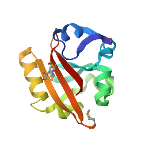
Initial events in the photocycle of photoactive yellow protein.

Kort, R.Hellingwerf, K.J.Ravelli, R.B.

(2004) J Biol Chem 279: 26417-26424

  • DOI: https://doi.org/10.1074/jbc.M311961200
  • Primary Citation of Related Structures:  
    1UWN, 1UWP

  • PubMed Abstract: 

    The light-induced isomerization of a double bond is the key event that allows the conversion of light energy into a structural change in photoactive proteins for many light-mediated biological processes, such as vision, photosynthesis, photomorphogenesis, and photo movement. Cofactors such as retinals, linear tetrapyrroles, and 4-hydroxy-cinnamic acid have been selected by nature that provide the essential double bond to transduce the light signal into a conformational change and eventually, a physiological response. Here we report the first events after light excitation of the latter chromophore, containing a single ethylene double bond, in a low temperature crystallographic study of the photoactive yellow protein. We measured experimental phases to overcome possible model bias, corrected for minimized radiation damage, and measured absorption spectra of crystals to analyze the photoproducts formed. The data show a mechanism for the light activation of photoactive yellow protein, where the energy to drive the remainder of the conformational changes is stored in a slightly strained but fully cis-chromophore configuration. In addition, our data indicate a role for backbone rearrangements during the very early structural events.


  • Organizational Affiliation

    Laboratory for Microbiology, Swammerdam Institute for Life Sciences, University of Amsterdam, Nieuwe Achtergracht 166, 1018 WV Amsterdam, The Netherlands. rkort@science.uva.nl


Macromolecules
Find similar proteins by:  (by identity cutoff)  |  3D Structure
Entity ID: 1
MoleculeChains Sequence LengthOrganismDetailsImage
PHOTOACTIVE YELLOW PROTEINA [auth X]125Halorhodospira halophilaMutation(s): 0 
UniProt
Find proteins for P16113 (Halorhodospira halophila)
Explore P16113 
Go to UniProtKB:  P16113
Entity Groups  
Sequence Clusters30% Identity50% Identity70% Identity90% Identity95% Identity100% Identity
UniProt GroupP16113
Sequence Annotations
Expand
  • Reference Sequence
Small Molecules
Ligands 2 Unique
IDChains Name / Formula / InChI Key2D Diagram3D Interactions
HC4
Query on HC4

Download Ideal Coordinates CCD File 
B [auth X]4'-HYDROXYCINNAMIC ACID
C9 H8 O3
NGSWKAQJJWESNS-ZZXKWVIFSA-N
SO4
Query on SO4

Download Ideal Coordinates CCD File 
C [auth X],
D [auth X]
SULFATE ION
O4 S
QAOWNCQODCNURD-UHFFFAOYSA-L
Modified Residues  1 Unique
IDChains TypeFormula2D DiagramParent
MSE
Query on MSE
A [auth X]L-PEPTIDE LINKINGC5 H11 N O2 SeMET
Experimental Data & Validation

Experimental Data

  • Method: X-RAY DIFFRACTION
  • Resolution: 1.20 Å
  • R-Value Free: 0.147 
  • R-Value Work: 0.124 
  • R-Value Observed: 0.125 
  • Space Group: P 63
Unit Cell:
Length ( Å )Angle ( ˚ )
a = 65.922α = 90
b = 65.922β = 90
c = 40.584γ = 120
Software Package:
Software NamePurpose
REFMACrefinement
XDSdata reduction
XSCALEdata scaling
SHARPphasing

Structure Validation

View Full Validation Report


Entry History 

Deposition Data

Revision History  (Full details and data files)

  • Version 1.0: 2004-03-25
    Type: Initial release
  • Version 1.1: 2011-07-13
    Changes: Atomic model, Version format compliance
  • Version 1.2: 2019-01-30
    Changes: Advisory, Data collection, Derived calculations, Experimental preparation
  • Version 1.3: 2019-02-27
    Changes: Data collection, Database references, Derived calculations, Experimental preparation