


Crystal Structure of a Mutant Escherichia Coli Thioredoxin with an Arginine Insertion in the Active Site
Nikkola, M., Langsetmo, K., Fuchs, J.A., Eklund, H.To be published.
Experimental Data Snapshot
wwPDB Validation 3D Report Full Report

Entity ID: 1 | |||||
|---|---|---|---|---|---|
| Molecule | Chains | Sequence Length | Organism | Details | Image |
| THIOREDOXIN | 109 | Escherichia coli | Mutation(s): 0 | 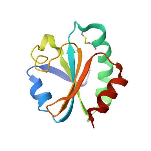 | |
UniProt | |||||
Find proteins for P0AA25 (Escherichia coli (strain K12)) Explore P0AA25 Go to UniProtKB: P0AA25 | |||||
Entity Groups | |||||
| Sequence Clusters | 30% Identity50% Identity70% Identity90% Identity95% Identity100% Identity | ||||
| UniProt Group | P0AA25 | ||||
Sequence AnnotationsExpand | |||||
| |||||
| Ligands 1 Unique | |||||
|---|---|---|---|---|---|
| ID | Chains | Name / Formula / InChI Key | 2D Diagram | 3D Interactions | |
| CU Query on CU | B [auth A] | COPPER (II) ION Cu JPVYNHNXODAKFH-UHFFFAOYSA-N |  | ||
| Length ( Å ) | Angle ( ˚ ) |
|---|---|
| a = 78.4 | α = 90 |
| b = 78.4 | β = 90 |
| c = 35.1 | γ = 120 |
| Software Name | Purpose |
|---|---|
| X-PLOR | model building |
| X-PLOR | refinement |
| X-PLOR | phasing |
RCSB PDB (citation) is hosted by
RCSB PDB is a member of the

