1T5T

Structure of the (SR)Ca2+-ATPase Ca2-E1-ADP:AlF4- form


Experimental Data Snapshot

  • Method: X-RAY DIFFRACTION
  • Resolution: 2.90 Å
  • R-Value Free: 0.293 
  • R-Value Work: 0.246 
  • R-Value Observed: 0.247 

wwPDB Validation   3D Report Full Report


Ligand Structure Quality Assessment 


This is version 1.3 of the entry. See complete history


Literature

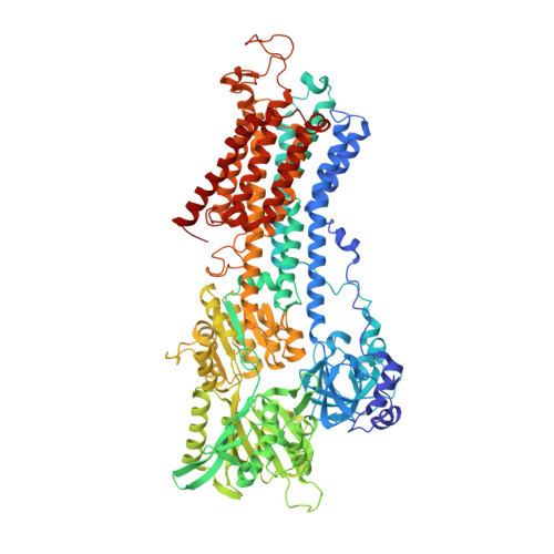
Phosphoryl transfer and calcium ion occlusion in the calcium pump.

Sorensen, T.L.Moller, J.V.Nissen, P.

(2004) Science 304: 1672-1675

  • DOI: https://doi.org/10.1126/science.1099366
  • Primary Citation of Related Structures:  
    1T5S, 1T5T

  • PubMed Abstract: 

    A tight coupling between adenosine triphosphate (ATP) hydrolysis and vectorial ion transport has to be maintained by ATP-consuming ion pumps. We report two crystal structures of Ca2+-bound sarco(endo)plasmic reticulum Ca2+-adenosine triphosphatase (SERCA) at 2.6 and 2.9 angstrom resolution in complex with (i) a nonhydrolyzable ATP analog [adenosine (beta-gamma methylene)-triphosphate] and (ii) adenosine diphosphate plus aluminum fluoride. SERCA reacts with ATP by an associative mechanism mediated by two Mg2+ ions to form an aspartyl-phosphorylated intermediate state (Ca2-E1 approximately P). The conformational changes that accompany the reaction with ATP pull the transmembrane helices 1 and 2 and close a cytosolic entrance for Ca2+, thereby preventing backflow before Ca2+ is released on the other side of the membrane.


  • Organizational Affiliation

    Department of Molecular Biology, University of Aarhus, Gustav Wieds Vej 10C, DK-8000 Aarhus C, Denmark.


Macromolecules
Find similar proteins by:  (by identity cutoff)  |  3D Structure
Entity ID: 1
MoleculeChains Sequence LengthOrganismDetailsImage
Sarcoplasmic/endoplasmic reticulum calcium ATPase 1 isoform SERCA1A994Oryctolagus cuniculusMutation(s): 0 
EC: 3.6.3.8
Membrane Entity: Yes 
UniProt
Find proteins for P04191 (Oryctolagus cuniculus)
Explore P04191 
Go to UniProtKB:  P04191
Entity Groups  
Sequence Clusters30% Identity50% Identity70% Identity90% Identity95% Identity100% Identity
UniProt GroupP04191
Sequence Annotations
Expand
  • Reference Sequence
Small Molecules
Ligands 5 Unique
IDChains Name / Formula / InChI Key2D Diagram3D Interactions
ADP
Query on ADP

Download Ideal Coordinates CCD File 
H [auth A]ADENOSINE-5'-DIPHOSPHATE
C10 H15 N5 O10 P2
XTWYTFMLZFPYCI-KQYNXXCUSA-N
ALF
Query on ALF

Download Ideal Coordinates CCD File 
B [auth A]TETRAFLUOROALUMINATE ION
Al F4
UYOMQIYKOOHAMK-UHFFFAOYSA-J
CA
Query on CA

Download Ideal Coordinates CCD File 
C [auth A],
D [auth A]
CALCIUM ION
Ca
BHPQYMZQTOCNFJ-UHFFFAOYSA-N
K
Query on K

Download Ideal Coordinates CCD File 
F [auth A]POTASSIUM ION
K
NPYPAHLBTDXSSS-UHFFFAOYSA-N
MG
Query on MG

Download Ideal Coordinates CCD File 
E [auth A],
G [auth A]
MAGNESIUM ION
Mg
JLVVSXFLKOJNIY-UHFFFAOYSA-N
Experimental Data & Validation

Experimental Data

  • Method: X-RAY DIFFRACTION
  • Resolution: 2.90 Å
  • R-Value Free: 0.293 
  • R-Value Work: 0.246 
  • R-Value Observed: 0.247 
  • Space Group: C 1 2 1
Unit Cell:
Length ( Å )Angle ( ˚ )
a = 162.477α = 90
b = 75.617β = 108.82
c = 151.651γ = 90
Software Package:
Software NamePurpose
REFMACrefinement
DENZOdata reduction
SCALEPACKdata scaling
CNSphasing

Structure Validation

View Full Validation Report


Ligand Structure Quality Assessment 


Entry History 

Deposition Data

Revision History  (Full details and data files)

  • Version 1.0: 2004-06-15
    Type: Initial release
  • Version 1.1: 2008-04-30
    Changes: Version format compliance
  • Version 1.2: 2011-07-13
    Changes: Advisory, Version format compliance
  • Version 1.3: 2024-02-14
    Changes: Data collection, Database references, Derived calculations