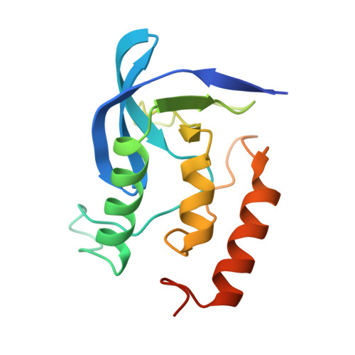
Engineering alternative beta-turn types in staphylococcal nuclease.
Hynes, T.R., Hodel, A., Fox, R.O.(1994) Biochemistry 33: 5021-5030
- PubMed: 8172877
- DOI: https://doi.org/10.1021/bi00183a004
- Primary Citation of Related Structures:
1SYC, 1SYD, 1SYE, 1SYF, 1SYG - PubMed Abstract:
We have refined the crystal structures of three point mutants of staphylococcal nuclease designed to favor alternative beta-turn types. Single amino acid substitutions were made in a type VIa beta-turn (residues 115-118; Tyr-Lys-Pro-Asn) containing a cis Lys 116-Pro 117 peptide bond. The mutations result in two new backbone conformations, a type I beta-turn for P117T and a type I' beta-turn for P117G and P117A. The P117G and P117A structures exhibit a dramatic difference in backbone conformation in the region of the mutation compared to the nuclease A structure such that the side chain of Lys 116 is reoriented to point into the nucleotide binding pocket. The distinct conformation observed for the nuclease A, P117G, and P117T beta-turn sequences agrees with correlations between beta-turn type and sequence identified from protein crystal structures. The P117A turn conformation provides an exception to these correlations. The results demonstrate that single residue changes can significantly alter backbone conformation, illustrating the process by which diversity in the structure of the protein surface can evolve on a conserved structural core, and suggest protein engineering applications in which the positioning as well as the identify of side chains can be modified to design new enzyme functions. Nuclease variants at the type VIa beta-turn site also allow the relationship between the amino acid sequence and beta-turn conformation to be examined in the context of an identical protein fold in crystallographic detail.
Organizational Affiliation:
Department of Molecular Biophysics and Biochemistry, Yale University, New Haven, Connecticut 06520.