1STH

TWO DISTINCTLY DIFFERENT METAL BINDING MODES ARE SEEN IN X-RAY CRYSTAL STRUCTURES OF STAPHYLOCOCCAL NUCLEASE-COBALT(II)-NUCLEOTIDE COMPLEXES


Experimental Data Snapshot

  • Method: X-RAY DIFFRACTION
  • Resolution: 1.85 Å
  • R-Value Work: 0.174 
  • R-Value Observed: 0.174 

wwPDB Validation   3D Report Full Report


Ligand Structure Quality Assessment 


This is version 1.3 of the entry. See complete history


Literature

X-ray crystal structures of staphylococcal nuclease complexed with the competitive inhibitor cobalt(II) and nucleotide.

Loll, P.J.Quirk, S.Lattman, E.E.Garavito, R.M.

(1995) Biochemistry 34: 4316-4324

  • DOI: https://doi.org/10.1021/bi00013a021
  • Primary Citation of Related Structures:  
    1STG, 1STH

  • PubMed Abstract: 

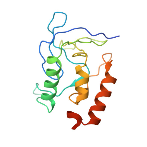
    Two crystal structures of ternary complexes of staphylococcal nuclease, cobalt(II), and the mononucleotide pdTp are reported. The first has been refined at 1.7 A to a crystallographic R value of 0.198; the second, determined from a crystal soaked for 9 months in a slightly different mother liquor than the first crystal, has been refined at 1.85 A to an R value of 0.174. In the first structure, the cobalt ion is displaced 1.94 A from the normal calcium position, and the active site is dominated by a salt bridge between Asp-21 and Lys-70 from a symmetry-related molecule in the crystal lattice. The Co2+ ion appears unable to displace this lysine; consequently, the metal is bound in a vestibular site adjacent to the calcium site. The metal-binding pocket in the second structure adopts a configuration similar to that of the calcium complex, with the cobalt ion binding only 0.36 A from the calcium position. However, an inner sphere water seen in the calcium structure is missing from this structure. The cobalt ion in the second structure appears to be loosely or transiently coordinated within the calcium binding pocket, as evidenced by the high value of its refined thermal factor. Loss of catalytic activity for cobalt(II)-substituted nuclease is perhaps due to its inability to bind this inner sphere water.


  • Organizational Affiliation

    Department of Biochemistry and Molecular Biology, University of Chicago, Illinois 60637, USA.


Macromolecules
Find similar proteins by:  (by identity cutoff)  |  3D Structure
Entity ID: 1
MoleculeChains Sequence LengthOrganismDetailsImage
STAPHYLOCOCCAL NUCLEASE149Staphylococcus aureusMutation(s): 0 
EC: 3.1.31.1
UniProt
Find proteins for P00644 (Staphylococcus aureus)
Explore P00644 
Go to UniProtKB:  P00644
Entity Groups  
Sequence Clusters30% Identity50% Identity70% Identity90% Identity95% Identity100% Identity
UniProt GroupP00644
Sequence Annotations
Expand
  • Reference Sequence
Small Molecules
Ligands 2 Unique
IDChains Name / Formula / InChI Key2D Diagram3D Interactions
THP
Query on THP

Download Ideal Coordinates CCD File 
C [auth A]THYMIDINE-3',5'-DIPHOSPHATE
C10 H16 N2 O11 P2
CSNCBOPUCJOHLS-XLPZGREQSA-N
CO
Query on CO

Download Ideal Coordinates CCD File 
B [auth A]COBALT (II) ION
Co
XLJKHNWPARRRJB-UHFFFAOYSA-N
Experimental Data & Validation

Experimental Data

  • Method: X-RAY DIFFRACTION
  • Resolution: 1.85 Å
  • R-Value Work: 0.174 
  • R-Value Observed: 0.174 
  • Space Group: P 41
Unit Cell:
Length ( Å )Angle ( ˚ )
a = 47.48α = 90
b = 47.48β = 90
c = 63.21γ = 90
Software Package:
Software NamePurpose
X-PLORmodel building
X-PLORrefinement
X-PLORphasing

Structure Validation

View Full Validation Report


Ligand Structure Quality Assessment 


Entry History 

Deposition Data

Revision History  (Full details and data files)

  • Version 1.0: 1995-02-27
    Type: Initial release
  • Version 1.1: 2008-03-24
    Changes: Version format compliance
  • Version 1.2: 2011-07-13
    Changes: Version format compliance
  • Version 1.3: 2024-02-14
    Changes: Data collection, Database references, Derived calculations, Other