1SRN

THE REFINED CRYSTAL STRUCTURE OF A FULLY ACTIVE SEMISYNTHETIC RIBONUCLEASE AT 1.8 ANGSTROMS RESOLUTION


Experimental Data Snapshot

  • Method: X-RAY DIFFRACTION
  • Resolution: 1.80 Å
  • R-Value Observed: 0.204 

wwPDB Validation   3D Report Full Report


This is version 1.2 of the entry. See complete history


Literature

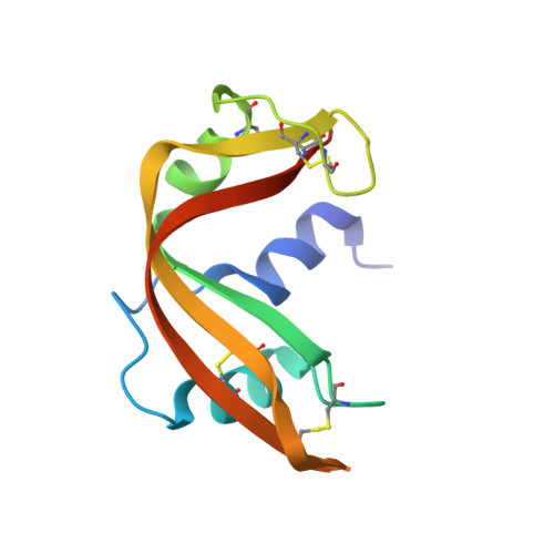
The refined crystal structure of a fully active semisynthetic ribonuclease at 1.8-A resolution.

Martin, P.D.Doscher, M.S.Edwards, B.F.

(1987) J Biol Chem 262: 15930-15938

  • DOI: https://doi.org/10.2210/pdb1srn/pdb
  • Primary Citation of Related Structures:  
    1SRN

  • PubMed Abstract: 

    A fully active, semisynthetic analog of bovine ribonuclease A, comprised of residues 1-118 of the molecule in a noncovalent complex with the synthetic peptide analog of residues 111-124, has been crystallized in space group P3(2)21 from a solution of 1.3 M ammonium sulfate and 3.0 M cesium chloride at pH 5.2. The crystallographic structure was determined by rotation and translation searches utilizing the coordinates for ribonuclease A reported by Wlodawer and Sjolin (Wlodawer, A., and Sjolin, L. (1983) Biochemistry 22, 2720-2728) and has been refined at 1.8-A resolution to an agreement factor of 0.204. Most of the structure of the semisynthetic enzyme closely resembles that found in ribonuclease A with the synthetic peptide replacing the C-terminal elements of the naturally occurring enzyme. No redundant structure is seen; residues 114-118 of the larger chain and residues 111-113 of the peptide do not appear in our map. The positions of those residues at or near the active site are very similar to, if not identical with, those previously reported by others, except for histidine 119, which occupies predominantly the B position seen as a minor site by Borkakoti et al. (Borkakoti, N., Moss, D. S., and Palmer, R. A. (1982) Acta Crystallogr. Sect. B Struct. Crystallogr. Cryst. Chem. 38,2210-2217) and not at all by Wlodawer and Sjolin (1983).


  • Organizational Affiliation

    Department of Biochemistry, Wayne State University School of Medicine, Detroit, Michigan 48201.


Macromolecules
Find similar proteins by:  (by identity cutoff)  |  3D Structure
Entity ID: 1
MoleculeChains Sequence LengthOrganismDetailsImage
RIBONUCLEASE A118Bos taurusMutation(s): 0 
EC: 3.1.27.5
UniProt
Find proteins for P61823 (Bos taurus)
Explore P61823 
Go to UniProtKB:  P61823
Entity Groups  
Sequence Clusters30% Identity50% Identity70% Identity90% Identity95% Identity100% Identity
UniProt GroupP61823
Sequence Annotations
Expand
  • Reference Sequence

Find similar proteins by:  Sequence   |   3D Structure  

Entity ID: 2
MoleculeChains Sequence LengthOrganismDetailsImage
RIBONUCLEASE A14Bos taurusMutation(s): 0 
UniProt
Find proteins for P61823 (Bos taurus)
Explore P61823 
Go to UniProtKB:  P61823
Entity Groups  
Sequence Clusters30% Identity50% Identity70% Identity90% Identity95% Identity100% Identity
UniProt GroupP61823
Sequence Annotations
Expand
  • Reference Sequence
Small Molecules
Ligands 1 Unique
IDChains Name / Formula / InChI Key2D Diagram3D Interactions
SO4
Query on SO4

Download Ideal Coordinates CCD File 
C [auth A]SULFATE ION
O4 S
QAOWNCQODCNURD-UHFFFAOYSA-L
Experimental Data & Validation

Experimental Data

  • Method: X-RAY DIFFRACTION
  • Resolution: 1.80 Å
  • R-Value Observed: 0.204 
  • Space Group: P 32 2 1
Unit Cell:
Length ( Å )Angle ( ˚ )
a = 67.68α = 90
b = 67.68β = 90
c = 65.03γ = 120
Software Package:
Software NamePurpose
PROLSQrefinement

Structure Validation

View Full Validation Report


Entry History 

Deposition Data

Revision History  (Full details and data files)

  • Version 1.0: 1990-10-15
    Type: Initial release
  • Version 1.1: 2008-03-24
    Changes: Version format compliance
  • Version 1.2: 2011-07-13
    Changes: Version format compliance