1SNC

THE CRYSTAL STRUCTURE OF THE TERNARY COMPLEX OF STAPHYLOCOCCAL NUCLEASE, CA2+, AND THE INHIBITOR PD*TP, REFINED AT 1.65 ANGSTROMS


Experimental Data Snapshot

  • Method: X-RAY DIFFRACTION
  • Resolution: 1.65 Å
  • R-Value Observed: 0.161 

wwPDB Validation   3D Report Full Report


Ligand Structure Quality Assessment 


This is version 1.4 of the entry. See complete history


Literature

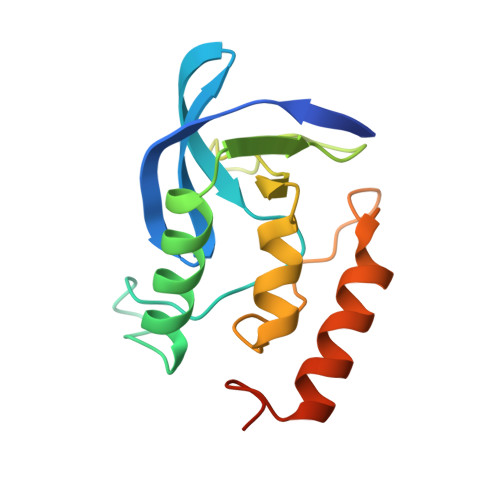
The crystal structure of the ternary complex of staphylococcal nuclease, Ca2+, and the inhibitor pdTp, refined at 1.65 A.

Loll, P.J.Lattman, E.E.

(1989) Proteins 5: 183-201

  • DOI: https://doi.org/10.1002/prot.340050302
  • Primary Citation of Related Structures:  
    1SNC

  • PubMed Abstract: 

    The structure of a complex of staphylococcal nuclease with Ca2+ and deoxythymidine 3',5'-bisphosphate (pdTp) has been refined by stereochemically restrained least-squares minimization to a crystallographic R value of 0.161 at 1.65 A resolution. The estimated root-mean-square (rms) error in the coordinates is 0.16 A. The final model comprises 1082 protein atoms, one calcium ion, the pdTp molecule, and 82 solvent water molecules; it displays an rms deviation from ideality of 0.017 A for bond distances and 1.8 degrees for bond angles. The mean distance between corresponding alpha carbons in the refined and unrefined structures is 0.6 A; we observe small but significant differences between the refined and unrefined models in the turn between residues 27 and 30, the loop between residues 44 and 50, the first helix, and the extended strand between residues 112 and 117 which forms part of the active site binding pocket. The details of the calcium liganding and solvent structure in the active site are clearly shown in the final electron density map. The structure of the catalytic site is consistent with the mechanism that has been proposed for this enzyme. However, we note that two lysines from a symmetry-related molecule in the crystal lattice may play an important role in determining the geometry of inhibitor binding, and that only one of the two required calcium ions is observed in the crystal structure; thus, caution is advised in extrapolating from the structure of the complex of enzyme and inhibitor to that of enzyme and substrate.


  • Organizational Affiliation

    Department of Biophysics, Johns Hopkins University School of Medicine, Baltimore, MD 21205.


Macromolecules
Find similar proteins by:  (by identity cutoff)  |  3D Structure
Entity ID: 1
MoleculeChains Sequence LengthOrganismDetailsImage
THERMONUCLEASE PRECURSOR149Staphylococcus aureusMutation(s): 0 
EC: 3.1.31.1
UniProt
Find proteins for P00644 (Staphylococcus aureus)
Explore P00644 
Go to UniProtKB:  P00644
Entity Groups  
Sequence Clusters30% Identity50% Identity70% Identity90% Identity95% Identity100% Identity
UniProt GroupP00644
Sequence Annotations
Expand
  • Reference Sequence
Small Molecules
Ligands 2 Unique
IDChains Name / Formula / InChI Key2D Diagram3D Interactions
THP
Query on THP

Download Ideal Coordinates CCD File 
C [auth A]THYMIDINE-3',5'-DIPHOSPHATE
C10 H16 N2 O11 P2
CSNCBOPUCJOHLS-XLPZGREQSA-N
CA
Query on CA

Download Ideal Coordinates CCD File 
B [auth A]CALCIUM ION
Ca
BHPQYMZQTOCNFJ-UHFFFAOYSA-N
Experimental Data & Validation

Experimental Data

  • Method: X-RAY DIFFRACTION
  • Resolution: 1.65 Å
  • R-Value Observed: 0.161 
  • Space Group: P 41
Unit Cell:
Length ( Å )Angle ( ˚ )
a = 48α = 90
b = 48β = 90
c = 63.5γ = 90
Software Package:
Software NamePurpose
PROLSQrefinement

Structure Validation

View Full Validation Report


Ligand Structure Quality Assessment 


Entry History 

Deposition Data

Revision History  (Full details and data files)

  • Version 1.0: 1990-10-15
    Type: Initial release
  • Version 1.1: 2008-03-03
    Changes: Version format compliance
  • Version 1.2: 2011-07-13
    Changes: Version format compliance
  • Version 1.3: 2017-11-29
    Changes: Derived calculations, Other
  • Version 1.4: 2024-02-14
    Changes: Data collection, Database references, Derived calculations