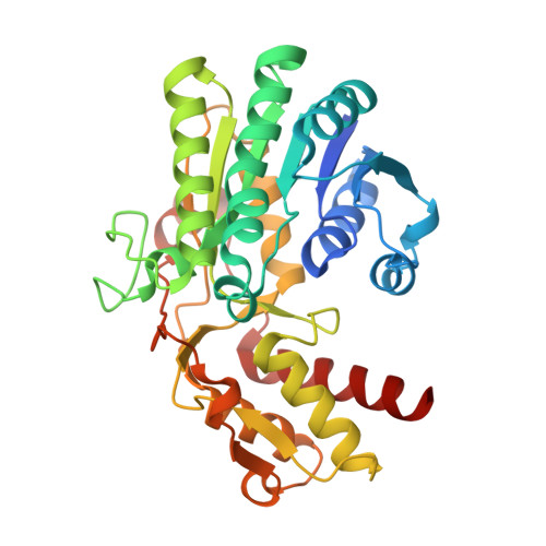
1RPN

Crystal Structure of GDP-D-mannose 4,6-dehydratase in complexes with GDP and NADPH


Experimental Data Snapshot

  • Method: X-RAY DIFFRACTION
  • Resolution: 2.15 Å
  • R-Value Free: 0.196 
  • R-Value Work: 0.174 
  • R-Value Observed: 0.176 

Starting Model: experimental
View more details

wwPDB Validation   3D Report Full Report


Ligand Structure Quality Assessment 


This is version 1.3 of the entry. See complete history


Literature

Crystal structure of a tetrameric GDP-D-mannose 4,6-dehydratase from a bacterial GDP-D-rhamnose biosynthetic pathway.

Webb, N.A.Mulichak, A.M.Lam, J.S.Rocchetta, H.L.Garavito, R.M.

(2004) Protein Sci 13: 529-539

  • DOI: https://doi.org/10.1110/ps.03393904
  • Primary Citation of Related Structures:  
    1RPN

  • PubMed Abstract: 

    d-Rhamnose is a rare 6-deoxy monosaccharide primarily found in the lipopolysaccharide of pathogenic bacteria, where it is involved in host-bacterium interactions and the establishment of infection. The biosynthesis of d-rhamnose proceeds through the conversion of GDP-d-mannose by GDP-d-mannose 4,6-dehydratase (GMD) to GDP-4-keto-6-deoxymannose, which is subsequently reduced to GDP-d-rhamnose by a reductase. We have determined the crystal structure of GMD from Pseudomonas aeruginosa in complex with NADPH and GDP. GMD belongs to the NDP-sugar modifying subfamily of the short-chain dehydrogenase/reductase (SDR) enzymes, all of which exhibit bidomain structures and a conserved catalytic triad (Tyr-XXX-Lys and Ser/Thr). Although most members of this enzyme subfamily display homodimeric structures, this bacterial GMD forms a tetramer in the same fashion as the plant MUR1 from Arabidopsis thaliana. The cofactor binding sites are adjoined across the tetramer interface, which brings the adenosyl phosphate moieties of the adjacent NADPH molecules to within 7 A of each other. A short peptide segment (Arg35-Arg43) stretches into the neighboring monomer, making not only protein-protein interactions but also hydrogen bonding interactions with the neighboring cofactor. The interface hydrogen bonds made by the Arg35-Arg43 segment are generally conserved in GMD and MUR1, and the interacting residues are highly conserved among the sequences of bacterial and eukaryotic GMDs. Outside of the Arg35-Arg43 segment, residues involved in tetrameric contacts are also quite conserved across different species. These observations suggest that a tetramer is the preferred, and perhaps functionally relevant, oligomeric state for most bacterial and eukaryotic GMDs.


  • Organizational Affiliation

    Department of Biochemistry and Molecular Biology, Michigan State University, East Lansing, MI 48824-1319, USA.


Macromolecules
Find similar proteins by:  (by identity cutoff)  |  3D Structure
Entity ID: 1
MoleculeChains Sequence LengthOrganismDetailsImage
GDP-mannose 4,6-dehydratase
A, B, C, D
335Pseudomonas aeruginosaMutation(s): 0 
Gene Names: GCAPA5453
EC: 4.2.1.47
UniProt
Find proteins for Q51366 (Pseudomonas aeruginosa (strain ATCC 15692 / DSM 22644 / CIP 104116 / JCM 14847 / LMG 12228 / 1C / PRS 101 / PAO1))
Explore Q51366 
Go to UniProtKB:  Q51366
Entity Groups  
Sequence Clusters30% Identity50% Identity70% Identity90% Identity95% Identity100% Identity
UniProt GroupQ51366
Sequence Annotations
Expand
  • Reference Sequence
Small Molecules
Ligands 2 Unique
IDChains Name / Formula / InChI Key2D Diagram3D Interactions
NDP
Query on NDP

Download Ideal Coordinates CCD File 
E [auth A],
G [auth B],
I [auth C],
K [auth D]
NADPH DIHYDRO-NICOTINAMIDE-ADENINE-DINUCLEOTIDE PHOSPHATE
C21 H30 N7 O17 P3
ACFIXJIJDZMPPO-NNYOXOHSSA-N
GDP
Query on GDP

Download Ideal Coordinates CCD File 
F [auth A],
H [auth B],
J [auth C],
L [auth D]
GUANOSINE-5'-DIPHOSPHATE
C10 H15 N5 O11 P2
QGWNDRXFNXRZMB-UUOKFMHZSA-N
Experimental Data & Validation

Experimental Data

  • Method: X-RAY DIFFRACTION
  • Resolution: 2.15 Å
  • R-Value Free: 0.196 
  • R-Value Work: 0.174 
  • R-Value Observed: 0.176 
  • Space Group: P 32 2 1
Unit Cell:
Length ( Å )Angle ( ˚ )
a = 125.73α = 90
b = 125.73β = 90
c = 220.025γ = 120
Software Package:
Software NamePurpose
DENZOdata reduction
SCALEPACKdata scaling
AMoREphasing
CNSrefinement

Structure Validation

View Full Validation Report


Ligand Structure Quality Assessment 


Entry History 

Deposition Data

Revision History  (Full details and data files)

  • Version 1.0: 2004-02-24
    Type: Initial release
  • Version 1.1: 2008-04-29
    Changes: Version format compliance
  • Version 1.2: 2011-07-13
    Changes: Derived calculations, Version format compliance
  • Version 1.3: 2023-08-23
    Changes: Data collection, Database references, Derived calculations, Refinement description