Refinement of the crystal structure of ribonuclease S. Comparison with and between the various ribonuclease A structures.
Kim, E.E., Varadarajan, R., Wyckoff, H.W., Richards, F.M.(1992) Biochemistry 31: 12304-12314
- PubMed: 1463719
- DOI: https://doi.org/10.1021/bi00164a004
- Primary Citation of Related Structures:
1RNU, 1RNV, 2RNS - PubMed Abstract:
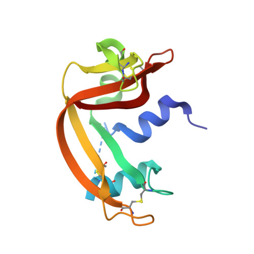
Ribonuclease S (RNase-S) is a complex that consists of two proteolytic fragments of bovine pancreatic ribonuclease A (RNase-A): the S-peptide (residues 1-20) and S-protein (residues 21-124). We have refined the crystal structures of three RNase-S complexes. The first two contain the full-length 20-residue S-peptide and were studied at pHs of 4.75 and 5.5. The third one consists of a truncated form of S-peptide (residues 1-15) and was studied at pH 4.75 as the reference structure for a series of mutant peptide complexes to be reported separately. Excluding residues 16-23 which are either missing (in the S15 complex) or disordered (in both S20 complexes), all three structures refined at 1.6-A resolution are identical within the estimated errors in the coordinates (0.048 A for the backbone atoms). The R-values, residual error, range from 17.4% to 18.6%. The final model of S20, pH 4.75, includes 1 sulfate and 84 water molecules. The side chains of 11 residues were modeled in two discrete conformations. The final structures were independent of the particular RNase-A or RNase-S used as a starting model. An extensive comparison with refined crystal structures of RNase-A reveals that the core of the molecule which is held together with extensive hydrogen bonds is in identical pattern in all cases. However, the loop regions vary from one structure to another and are often characterized by high B-factors. The pattern of thermal parameters appears to be dependent on crystal packing and correlates well with the accessibility calculated in the crystal. Gln60 is a conserved residue in all sequences known to date for this class of ribonucleases. However, it is the only residue that is clearly defined in an unfavorable position (phi = -100 degrees, psi = -130 degrees) on the Ramachandran plot. The origin of the substantial differences between RNase-A and RNase-S in stability to both acid and temperature denaturation and in susceptibility to proteolysis at neutral pH is not obvious in our visual comparison of these two structures.
Organizational Affiliation:
Department of Molecular Biophysics and Biochemistry, Yale University, New Haven, Connecticut 06511.