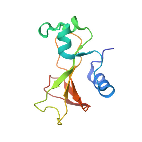
1RNB

CRYSTAL STRUCTURE OF A BARNASE-D(*GP*C) COMPLEX AT 1.9 ANGSTROMS RESOLUTION


Experimental Data Snapshot

  • Method: X-RAY DIFFRACTION
  • Resolution: 1.90 Å
  • R-Value Work: 0.214 

wwPDB Validation   3D Report Full Report


This is version 1.3 of the entry. See complete history


Literature

Crystal structure of a barnase-d(GpC) complex at 1.9 A resolution.

Baudet, S.Janin, J.

(1991) J Mol Biol 219: 123-132

  • DOI: https://doi.org/10.1016/0022-2836(91)90862-z
  • Primary Citation of Related Structures:  
    1RNB

  • PubMed Abstract: 

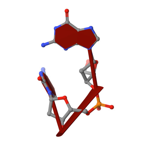
    The ribonuclease excreted by Bacillus amyloliquefaciens, Barnase, was co-crystallized with the deoxy-dinucleotide d(GpC). The crystal structure was determined by molecular replacement from a model of free Barnase previously derived by Mauguen et al. Refinement was carried out using data to 1.9 A resolution. The final model, which has a crystallographic R factor of 22%, includes 869 protein atoms, 38 atoms from d(GpC), a sulfate ion and 73 water molecules. Only minor differences from free Barnase are seen in the protein moiety, the root-mean-square C alpha movement being 0.45 A. The dinucleotide has a folded conformation. It is located near the active site of the enzyme, but outside the protein molecule and making crystal packing contacts with neighboring molecules. The guanine base is stacked on the imidazole ring of active site His102, rather than binding to the so-called recognition loop as it does in other complexes of guanine nucleotides with microbial nucleases. The deoxyguanosine is syn, with the sugar ring in C-2'-endo conformation; the deoxycytidine is anti and C-4'-exo. In addition to the stacking interaction, His102 hydrogen bonds to the free 5' hydroxyl, which is located near the position where the 3' phosphate group is found in other inhibitors of microbial ribonucleases. While the mode of binding observed with d(GpC) and Barnase would be non-productive for a dinucleotide substrate, it may define a site for the nucleotide product on the 3' side of the hydrolyzed bond.


  • Organizational Affiliation

    Laboratoire de Biologie Physicochimique-CNRS U.A. 1131, Université Paris-Sud, Orsay, France.


Macromolecules

Find similar proteins by:  (by identity cutoff)  |  3D Structure
Entity ID: 2
MoleculeChains Sequence LengthOrganismDetailsImage
BARNASEB [auth A]110Bacillus amyloliquefaciensMutation(s): 0 
UniProt
Find proteins for P00648 (Bacillus amyloliquefaciens)
Explore P00648 
Go to UniProtKB:  P00648
Entity Groups  
Sequence Clusters30% Identity50% Identity70% Identity90% Identity95% Identity100% Identity
UniProt GroupP00648
Sequence Annotations
Expand
  • Reference Sequence

Find similar nucleic acids by:  Sequence   |   3D Structure  

Entity ID: 1
MoleculeChains LengthOrganismImage
DNA (5'-D(*GP*C)-3')A [auth C]2N/A
Sequence Annotations
Expand
  • Reference Sequence
Small Molecules
Ligands 1 Unique
IDChains Name / Formula / InChI Key2D Diagram3D Interactions
SO4
Query on SO4

Download Ideal Coordinates CCD File 
C
SULFATE ION
O4 S
QAOWNCQODCNURD-UHFFFAOYSA-L
Experimental Data & Validation

Experimental Data

  • Method: X-RAY DIFFRACTION
  • Resolution: 1.90 Å
  • R-Value Work: 0.214 
  • Space Group: P 31 2 1
Unit Cell:
Length ( Å )Angle ( ˚ )
a = 57.97α = 90
b = 57.97β = 90
c = 85.37γ = 120
Software Package:
Software NamePurpose
PROLSQrefinement

Structure Validation

View Full Validation Report


Entry History 

Deposition Data

Revision History  (Full details and data files)

  • Version 1.0: 1992-07-15
    Type: Initial release
  • Version 1.1: 2008-05-22
    Changes: Version format compliance
  • Version 1.2: 2011-07-13
    Changes: Version format compliance
  • Version 1.3: 2024-02-14
    Changes: Data collection, Database references, Derived calculations, Other