1RLS

CRYSTAL STRUCTURE OF RNASE T1 COMPLEXED WITH THE PRODUCT NUCLEOTIDE 3'-GMP. STRUCTURAL EVIDENCE FOR DIRECT INTERACTION OF HISTIDINE 40 AND GLUTAMIC ACID 58 WITH THE 2'-HYDROXYL GROUP OF RIBOSE


Experimental Data Snapshot

  • Method: X-RAY DIFFRACTION
  • Resolution: 1.90 Å
  • R-Value Observed: 0.167 

wwPDB Validation   3D Report Full Report


This is version 1.3 of the entry. See complete history


Literature

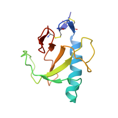
Crystal structure of RNase T1 complexed with the product nucleotide 3'-GMP. Structural evidence for direct interaction of histidine 40 and glutamic acid 58 with the 2'-hydroxyl group of the ribose.

Gohda, K.Oka, K.Tomita, K.Hakoshima, T.

(1994) J Biol Chem 269: 17531-17536

  • DOI: https://doi.org/10.2210/pdb1rls/pdb
  • Primary Citation of Related Structures:  
    1RLS

  • PubMed Abstract: 

    The crystal structure of RNase T1 complexed with 3'-GMP has been determined. The glycosyl conformation of 3'-GMP is in the syn conformation, and the ribose adopts the O4'-endo pucker. This observed pucker is different from that in any complex structures of RNase T1. In the present complex, this energetically unfavorable conformation is stabilized by the water molecule with the bridged hydrogen bonds between the O2' and the O3' atoms of the ribose. The guanine base is recognized in the same manner as observed in the complex of 2'-GMP. The 2'-hydroxyl group of the ribose shows a tight hydrogen bond to both His-40 and Glu-58 with the suitable geometry for the proton transfer. These hydrogen bonds suggest that the two residues can participate directly in the proton transfer. His-92 is hydrogen bonded to two the proton transfer. His-92 is hydrogen bonded to two oxygen atoms of the phosphate group. Based on the geometry in the active site, the O1P atom may correspond to the O5' atom of the leaving nucleotide in the phosphoryl transfer or a water molecule as a nucleophile in the hydrolysis reaction. In the present complex, the conformations of the 3'-GMP molecule and the side chains of the catalytic residues would be represented as the conformation before the phosphoryl transfer reaction and/or after the hydrolysis reaction.


  • Organizational Affiliation

    Faculty of Pharmaceutical Sciences, Osaka University, Japan.


Macromolecules
Find similar proteins by:  (by identity cutoff)  |  3D Structure
Entity ID: 1
MoleculeChains Sequence LengthOrganismDetailsImage
RIBONUCLEASE T1104Aspergillus oryzaeMutation(s): 0 
EC: 3.1.27.3
UniProt
Find proteins for P00651 (Aspergillus oryzae (strain ATCC 42149 / RIB 40))
Explore P00651 
Go to UniProtKB:  P00651
Entity Groups  
Sequence Clusters30% Identity50% Identity70% Identity90% Identity95% Identity100% Identity
UniProt GroupP00651
Sequence Annotations
Expand
  • Reference Sequence
Small Molecules
Ligands 2 Unique
IDChains Name / Formula / InChI Key2D Diagram3D Interactions
3GP
Query on 3GP

Download Ideal Coordinates CCD File 
C [auth A]GUANOSINE-3'-MONOPHOSPHATE
C10 H14 N5 O8 P
ZDPUTNZENXVHJC-UUOKFMHZSA-N
CA
Query on CA

Download Ideal Coordinates CCD File 
B [auth A]CALCIUM ION
Ca
BHPQYMZQTOCNFJ-UHFFFAOYSA-N
Experimental Data & Validation

Experimental Data

  • Method: X-RAY DIFFRACTION
  • Resolution: 1.90 Å
  • R-Value Observed: 0.167 
  • Space Group: P 21 21 21
Unit Cell:
Length ( Å )Angle ( ˚ )
a = 48.14α = 90
b = 51.06β = 90
c = 40.38γ = 90
Software Package:
Software NamePurpose
X-PLORmodel building
PROFFTrefinement
X-PLORrefinement
X-PLORphasing

Structure Validation

View Full Validation Report


Entry History 

Deposition Data

Revision History  (Full details and data files)

  • Version 1.0: 1994-12-20
    Type: Initial release
  • Version 1.1: 2008-03-24
    Changes: Version format compliance
  • Version 1.2: 2011-07-13
    Changes: Version format compliance
  • Version 1.3: 2017-11-29
    Changes: Derived calculations, Other