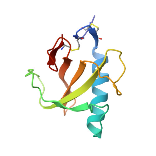
The complex between ribonuclease T1 and 3'GMP suggests geometry of enzymic reaction path. An X-ray study.
Heydenreich, A., Koellner, G., Choe, H.W., Cordes, F., Kisker, C., Schindelin, H., Adamiak, R., Hahn, U., Saenger, W.(1993) Eur J Biochem 218: 1005-1012
- PubMed: 8281918
- DOI: https://doi.org/10.1111/j.1432-1033.1993.tb18459.x
- Primary Citation of Related Structures:
1RGC - PubMed Abstract:
The crystal structure of the complex between ribonuclease T1 and 3'GMP suggests that (a) a substrate GpN is bound to the active site of ribonuclease T1 in a conformation that actively supports the catalytic process, (b) the reaction occurs in an in-line process, (c) His40 N epsilon H+ activates O2'-H, (d) Glu58 carboxylate acts as base and His92 N epsilon H+ as acid in a general acid-base catalysis. The crystals have the monoclinic space group P2(1), a = 4.968 nm, b = 4.833 nm, c = 4.048 nm, beta = 90.62 degrees with two molecules in the asymmetric unit. The structure was determined by molecular replacement and refined to R = 15.3% with 11,338 data > or = 1 sigma (Fo) in the resolution range 1.0-0.2 nm; this includes 180 water molecules and two Ca2+. The structure of ribonuclease T1 is as previously observed. 3'GMP is bound in syn conformation; guanine is located in the specific recognition site, the ribose adopts C4'-exo puckering, the ribose phosphate is extended with torsion angle epsilon in trans. The O2'-H group is activated by accepting and donating hydrogen bonds from His40 N epsilon H+ and to Glu58 O epsilon 1; the phosphate is hydrogen bonded to Glu58 O epsilon 2H, Arg77 N epsilon H+ and N eta 2H+, Tyr38 O eta H, His92 N eta H+. The conformation of ribose phosphate is such that O2' is at a distance of 0.31 nm from phosphorus, and opposite the P-OP3 bond which accepts a hydrogen bond from His92 N epsilon H+; we infer from a model building study that this bond is equivalent to the scissile P-O5' in a substrate GpN.
Organizational Affiliation:
Institut für Kristallographie, Freie Universität Berlin, Germany.