1Q5I

Crystal structure of bacteriorhodopsin mutant P186A crystallized from bicelles


Experimental Data Snapshot

  • Method: X-RAY DIFFRACTION
  • Resolution: 2.30 Å
  • R-Value Free: 0.256 
  • R-Value Work: 0.194 
  • R-Value Observed: 0.198 

wwPDB Validation   3D Report Full Report


This is version 1.5 of the entry. See complete history


Literature

The evolution of transmembrane helix kinks and the structural diversity of G protein-coupled receptors.

Yohannan, S.Faham, S.Yang, D.Whitelegge, J.P.Bowie, J.U.

(2004) Proc Natl Acad Sci U S A 101: 959-963

  • DOI: https://doi.org/10.1073/pnas.0306077101
  • Primary Citation of Related Structures:  
    1Q5I, 1Q5J

  • PubMed Abstract: 

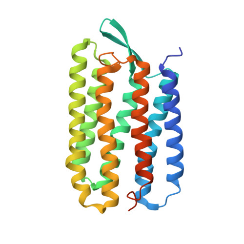
    One of the hallmarks of membrane protein structure is the high frequency of transmembrane helix kinks, which commonly occur at proline residues. Because the proline side chain usually precludes normal helix geometry, it is reasonable to expect that proline residues generate these kinks. We observe, however, that the three prolines in bacteriorhodopsin transmembrane helices can be changed to alanine with little structural consequences. This finding leads to a conundrum: if proline is not required for helix bending, why are prolines commonly present at bends in transmembrane helices? We propose an evolutionary hypothesis in which a mutation to proline initially induces the kink. The resulting packing defects are later repaired by further mutation, thereby locking the kink in the structure. Thus, most prolines in extant proteins can be removed without major structural consequences. We further propose that nonproline kinks are places where vestigial prolines were later removed during evolution. Consistent with this hypothesis, at 14 of 17 nonproline kinks in membrane proteins of known structure, we find prolines in homologous sequences. Our analysis allows us to predict kink positions with >90% reliability. Kink prediction indicates that different G protein-coupled receptor proteins have different kink patterns and therefore different structures.


  • Organizational Affiliation

    Department of Chemistry and Biochemistry, UCLA-DOE Institute for Genomics and Proteomics, Molecular Biology Institute, University of California, Los Angeles, CA 90095, USA.


Macromolecules
Find similar proteins by:  (by identity cutoff)  |  3D Structure
Entity ID: 1
MoleculeChains Sequence LengthOrganismDetailsImage
Bacteriorhodopsin
A, B
249Halobacterium salinarumMutation(s): 1 
Gene Names: BOP OR VNG1467G
Membrane Entity: Yes 
UniProt
Find proteins for P02945 (Halobacterium salinarum (strain ATCC 700922 / JCM 11081 / NRC-1))
Explore P02945 
Go to UniProtKB:  P02945
Entity Groups  
Sequence Clusters30% Identity50% Identity70% Identity90% Identity95% Identity100% Identity
UniProt GroupP02945
Sequence Annotations
Expand
  • Reference Sequence
Small Molecules
Ligands 1 Unique
IDChains Name / Formula / InChI Key2D Diagram3D Interactions
RET
Query on RET

Download Ideal Coordinates CCD File 
C [auth A],
D [auth B]
RETINAL
C20 H28 O
NCYCYZXNIZJOKI-OVSJKPMPSA-N
Experimental Data & Validation

Experimental Data

  • Method: X-RAY DIFFRACTION
  • Resolution: 2.30 Å
  • R-Value Free: 0.256 
  • R-Value Work: 0.194 
  • R-Value Observed: 0.198 
  • Space Group: P 1 21 1
Unit Cell:
Length ( Å )Angle ( ˚ )
a = 44.629α = 90
b = 108.721β = 113.46
c = 56.093γ = 90
Software Package:
Software NamePurpose
DENZOdata reduction
SCALEPACKdata scaling
CNSrefinement
CNSphasing

Structure Validation

View Full Validation Report


Entry History 

Deposition Data

Revision History  (Full details and data files)

  • Version 1.0: 2004-01-06
    Type: Initial release
  • Version 1.1: 2008-04-29
    Changes: Version format compliance
  • Version 1.2: 2011-07-13
    Changes: Version format compliance
  • Version 1.3: 2018-01-31
    Changes: Experimental preparation
  • Version 1.4: 2021-10-27
    Changes: Database references, Derived calculations
  • Version 1.5: 2023-08-16
    Changes: Data collection, Refinement description