1NZV

Crystal Structure of Src SH2 domain bound to doubly phosphorylated peptide PQpYIpYVPA


Experimental Data Snapshot

  • Method: X-RAY DIFFRACTION
  • Resolution: 2.10 Å
  • R-Value Free: 0.271 
  • R-Value Work: 0.224 

wwPDB Validation   3D Report Full Report


This is version 1.5 of the entry. See complete history


Literature

Structural and Thermodynamic Basis for the Interaction of the Src SH2 domain with the Activated form of the PDGF beta-receptor

Lubman, O.Y.Waksman, G.

(2003) J Mol Biol 328: 655-668

  • DOI: https://doi.org/10.1016/s0022-2836(03)00344-9
  • Primary Citation of Related Structures:  
    1NZL, 1NZV

  • PubMed Abstract: 

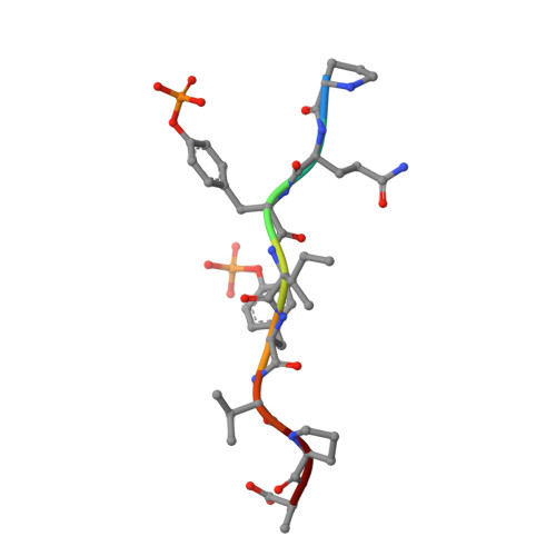
    Recruitment of the Src kinase to the activated form of the platelet-derived growth factor (PDGF) receptor involves recognition of a unique sequence motif in the juxtamembrane region of the receptor by the Src homology 2 (SH2) domain of the enzyme. This motif contains two phosphotyrosine residues separated by one residue (sequence pYIpYV where pY indicates a phosphotyrosine). Here, we provide the thermodynamic and structural basis for the binding of this motif by the Src SH2 domain. We show that the second phosphorylation event increases the free energy window for specific interaction and that the physiological target is exquisitely designed for the task of recruiting specifically an SH2 domain which otherwise demonstrates very little intrinsic ability to discriminate sequences C-terminal to the first phosphorylation event. Surprisingly, we show that water plays a role in the recognition process.


  • Organizational Affiliation

    Department of Biochemistry and Molecular Biophysics, School of Medicine, Washington University, 660 South Euclid Avenue, Saint Louis, MO 63110, USA.


Macromolecules
Find similar proteins by:  (by identity cutoff)  |  3D Structure
Entity ID: 1
MoleculeChains Sequence LengthOrganismDetailsImage
Tyrosine-protein kinase transforming protein SRC
A, B
103Rous sarcoma virus (strain Schmidt-Ruppin)Mutation(s): 0 
Gene Names: V-SRC
EC: 2.7.1.112
UniProt
Find proteins for P00524 (Rous sarcoma virus subgroup A (strain Schmidt-Ruppin))
Explore P00524 
Go to UniProtKB:  P00524
Entity Groups  
Sequence Clusters30% Identity50% Identity70% Identity90% Identity95% Identity100% Identity
UniProt GroupP00524
Sequence Annotations
Expand
  • Reference Sequence

Find similar proteins by:  Sequence   |   3D Structure  

Entity ID: 2
MoleculeChains Sequence LengthOrganismDetailsImage
Doubly phosphorylated peptide PQpYIpYVPA8N/AMutation(s): 0 
Sequence Annotations
Expand
  • Reference Sequence
Small Molecules
Modified Residues  1 Unique
IDChains TypeFormula2D DiagramParent
PTR
Query on PTR
C
L-PEPTIDE LINKINGC9 H12 N O6 PTYR
Binding Affinity Annotations 
IDSourceBinding Affinity
PTR BindingDB:  1NZV -TΔS: -1.81e+1 (kJ/mol) from 1 assay(s)
ΔG: -2.14e+1 (kJ/mol) from 1 assay(s)
Experimental Data & Validation

Experimental Data

  • Method: X-RAY DIFFRACTION
  • Resolution: 2.10 Å
  • R-Value Free: 0.271 
  • R-Value Work: 0.224 
  • Space Group: P 21 21 2
Unit Cell:
Length ( Å )Angle ( ˚ )
a = 100.256α = 90
b = 67.727β = 90
c = 29.636γ = 90
Software Package:
Software NamePurpose
CNSrefinement
DENZOdata reduction
SCALEPACKdata scaling
CNSphasing

Structure Validation

View Full Validation Report


Entry History 

Deposition Data

Revision History  (Full details and data files)

  • Version 1.0: 2003-04-22
    Type: Initial release
  • Version 1.1: 2008-04-29
    Changes: Version format compliance
  • Version 1.2: 2011-07-13
    Changes: Version format compliance
  • Version 1.3: 2014-04-30
    Changes: Data collection
  • Version 1.4: 2023-08-16
    Changes: Data collection, Database references, Derived calculations, Refinement description
  • Version 1.5: 2023-11-15
    Changes: Data collection