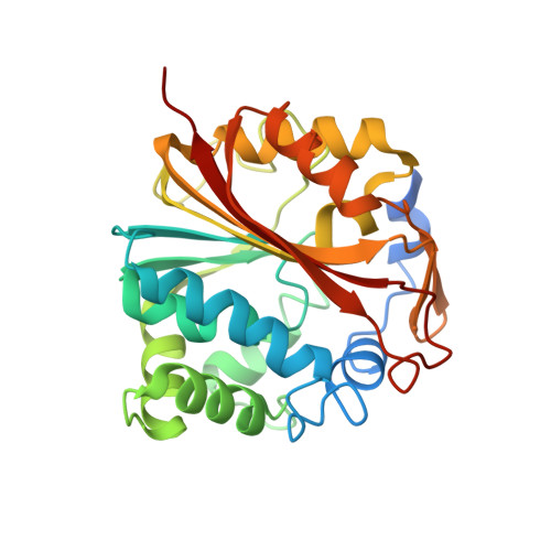
1N7J

The structure of Phenylethanolamine N-methyltransferase in complex with S-adenosylhomocysteine and an iodinated inhibitor


Experimental Data Snapshot

  • Method: X-RAY DIFFRACTION
  • Resolution: 2.70 Å
  • R-Value Free: 0.268 
  • R-Value Work: 0.232 
  • R-Value Observed: 0.232 

Starting Model: experimental
View more details

wwPDB Validation   3D Report Full Report


Ligand Structure Quality Assessment 


This is version 1.3 of the entry. See complete history


Literature

Molecular recognition of sub-micromolar inhibitors by the epinephrine-synthesizing enzyme phenylethanolamine N-methyltransferase.

McMillan, F.M.Archbold, J.McLeish, M.J.Caine, J.M.Criscione, K.R.Grunewald, G.L.Martin, J.L.

(2004) J Med Chem 47: 37-44

  • DOI: https://doi.org/10.1021/jm0205752
  • Primary Citation of Related Structures:  
    1N7I, 1N7J

  • PubMed Abstract: 

    The crystal structures of human phenylethanolamine N-methyltransferase in complex with S-adenosyl-l-homocysteine (7, AdoHcy) and either 7-iodo-1,2,3,4-tetrahydroisoquinoline (2) or 8,9-dichloro-2,3,4,5-tetrahydro-1H-2-benzazepine (3, LY134046) were determined and compared with the structure of the enzyme complex with 7 and 7-aminosulfonyl-1,2,3,4-tetrahydroisoquinoline (1, SK&F 29661). The enzyme is able to accommodate a variety of chemically disparate functional groups on the aromatic ring of the inhibitors through adaptation of the binding pocket for this substituent and by subtle adjustments of the orientation of the inhibitors within the relatively planar binding site. In addition, the interactions formed by the amine nitrogen of all three inhibitors reinforce the hypothesis that this functional group mimics the beta-hydroxyl of norepinephrine rather than the amine. These studies provide further clues for the development of improved inhibitors for use as pharmacological probes.


  • Organizational Affiliation

    Centre for Drug Design and Development and Special Research Centre for Functional and Applied Genomics, Institute for Molecular Bioscience, University of Queensland, Brisbane QLD 4072, Australia.


Macromolecules
Find similar proteins by:  (by identity cutoff)  |  3D Structure
Entity ID: 1
MoleculeChains Sequence LengthOrganismDetailsImage
Phenylethanolamine N-methyltransferase
A, B
282Homo sapiensMutation(s): 0 
Gene Names: PNMT OR PENT
EC: 2.1.1.28
UniProt & NIH Common Fund Data Resources
Find proteins for P11086 (Homo sapiens)
Explore P11086 
Go to UniProtKB:  P11086
PHAROS:  P11086
GTEx:  ENSG00000141744 
Entity Groups  
Sequence Clusters30% Identity50% Identity70% Identity90% Identity95% Identity100% Identity
UniProt GroupP11086
Sequence Annotations
Expand
  • Reference Sequence
Small Molecules
Binding Affinity Annotations 
IDSourceBinding Affinity
IDI BindingDB:  1N7J Ki: min: 40, max: 93 (nM) from 2 assay(s)
PDBBind:  1N7J Ki: 370 (nM) from 1 assay(s)
SAH BindingDB:  1N7J Ki: 1.40e+4 (nM) from 1 assay(s)
Experimental Data & Validation

Experimental Data

  • Method: X-RAY DIFFRACTION
  • Resolution: 2.70 Å
  • R-Value Free: 0.268 
  • R-Value Work: 0.232 
  • R-Value Observed: 0.232 
  • Space Group: P 43 21 2
Unit Cell:
Length ( Å )Angle ( ˚ )
a = 94.74α = 90
b = 94.74β = 90
c = 186.9γ = 90
Software Package:
Software NamePurpose
CNSrefinement
DENZOdata reduction
SCALEPACKdata scaling
CNSphasing

Structure Validation

View Full Validation Report


Ligand Structure Quality Assessment 


Entry History 

Deposition Data

Revision History  (Full details and data files)

  • Version 1.0: 2003-12-23
    Type: Initial release
  • Version 1.1: 2008-04-28
    Changes: Version format compliance
  • Version 1.2: 2011-07-13
    Changes: Version format compliance
  • Version 1.3: 2024-02-14
    Changes: Data collection, Database references, Derived calculations, Refinement description