1MVT

Analysis of Two Polymorphic Forms of a Pyrido[2,3-d]pyrimidine N9-C10 Reverse-Bridge Antifolate Binary Complex with Human Dihydrofolate Reductase


Experimental Data Snapshot

  • Method: X-RAY DIFFRACTION
  • Resolution: 1.80 Å
  • R-Value Work: 0.194 

wwPDB Validation   3D Report Full Report


Ligand Structure Quality Assessment 


This is version 1.4 of the entry. See complete history


Literature

Analysis of two polymorphic forms of a pyrido[2,3-d]pyrimidine N9-C10 reversed-bridge antifolate binary complex with human dihydrofolate reductase.

Cody, V.Galitsky, N.Luft, J.R.Pangborn, W.Gangjee, A.

(2003) Acta Crystallogr D Biol Crystallogr 59: 654-661

  • DOI: https://doi.org/10.1107/s0907444903001951
  • Primary Citation of Related Structures:  
    1MVS, 1MVT

  • PubMed Abstract: 

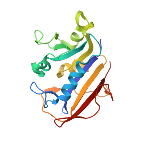
    The results of the crystal structure determination of human dihydrofolate reductase (hDHFR) as a binary complex with the potent N9-C10 reversed-bridge antifolate inhibitor 2,4-diamino-6-[N-(3',4',5'-trimethoxybenzyl)-N-methylamino]pyrido[2,3-d]pyrimidine (1) are reported for two independent polymorphic rhombohedral R3 lattices [R3(1) and R3(2)]. Data from these two crystal forms were refined to 1.90 A resolution for complex R3(1), with R = 0.186 for 9689 data, and to 1.80 A resolution for complex R3(2), with R = 0.194 for 13 305 data. Changes in the loop geometry between the two structures reflects contact differences in the packing environments in the two R3 lattices. The largest changes (between 0.5 and 1.7 A) are observed for the loop regions encompassing residues 16-25, 40-48, 81-89, 99-108, 143-148 and 161-169. Comparison of the intermolecular contacts of these loops reveals that the R3(2) lattice is more tightly packed, as reflected in its smaller V(M) value and smaller solvent content. The conformation of inhibitor (1) is similar in both structures and the N9-C10 bridge geometry is more similar to that observed for the normal C9-N10 bridge of trimetrexate (TMQ) than to the other N9-C10 reversed-bridge antifolates previously reported. The effect of the N9-C10 reversed-bridge geometry is to distort the bridge from coplanarity with the pyrido[2,3-d]pyrimidine ring system and to twist the C10 methylene conformation towards a gauche conformation. This also influences the conformation of the methoxybenzyl ring, moving it away from a trans position and placing the 5'-methoxy group deeper within the hydrophobic pocket made by Leu60, Pro61 and Asn64 of the hDHFR active site.


  • Organizational Affiliation

    Hauptman-Woodward Medical Research Institute Inc., Buffalo, NY 14203, USA. cody@hwi.buffalo.edu


Macromolecules
Find similar proteins by:  (by identity cutoff)  |  3D Structure
Entity ID: 1
MoleculeChains Sequence LengthOrganismDetailsImage
Dihydrofolate Reductase187Homo sapiensMutation(s): 0 
EC: 1.5.1.3
UniProt & NIH Common Fund Data Resources
Find proteins for P00374 (Homo sapiens)
Explore P00374 
Go to UniProtKB:  P00374
PHAROS:  P00374
GTEx:  ENSG00000228716 
Entity Groups  
Sequence Clusters30% Identity50% Identity70% Identity90% Identity95% Identity100% Identity
UniProt GroupP00374
Sequence Annotations
Expand
  • Reference Sequence
Small Molecules
Ligands 2 Unique
IDChains Name / Formula / InChI Key2D Diagram3D Interactions
DTM
Query on DTM

Download Ideal Coordinates CCD File 
D [auth A]2,4-DIAMINO-6-[N-(3',4',5'-TRIMETHOXYBENZYL)-N-METHYLAMINO]PYRIDO[2,3-D]PYRIMIDINE
C18 H22 N6 O3
PUOZHLHNKHRTOW-UHFFFAOYSA-N
SO4
Query on SO4

Download Ideal Coordinates CCD File 
B [auth A],
C [auth A]
SULFATE ION
O4 S
QAOWNCQODCNURD-UHFFFAOYSA-L
Binding Affinity Annotations 
IDSourceBinding Affinity
DTM BindingDB:  1MVT IC50: 33 (nM) from 1 assay(s)
Experimental Data & Validation

Experimental Data

  • Method: X-RAY DIFFRACTION
  • Resolution: 1.80 Å
  • R-Value Work: 0.194 
  • Space Group: H 3
Unit Cell:
Length ( Å )Angle ( ˚ )
a = 106.78α = 90
b = 106.78β = 90
c = 43.82γ = 120
Software Package:
Software NamePurpose
DENZOdata reduction
SCALEPACKdata scaling
PROTEINmodel building
PROLSQrefinement
PROTEINphasing

Structure Validation

View Full Validation Report


Ligand Structure Quality Assessment 


Entry History 

Deposition Data

Revision History  (Full details and data files)

  • Version 1.0: 2003-04-01
    Type: Initial release
  • Version 1.1: 2008-04-28
    Changes: Version format compliance
  • Version 1.2: 2011-07-13
    Changes: Version format compliance
  • Version 1.3: 2018-02-14
    Changes: Experimental preparation
  • Version 1.4: 2024-02-14
    Changes: Data collection, Database references, Derived calculations, Refinement description