A multigeneration analysis of cytochrome b(562) redox variants: evolutionary strategies for modulating redox potential revealed using a library approach.
Springs, S.L., Bass, S.E., Bowman, G., Nodelman, I., Schutt, C.E., McLendon, G.L.(2002) Biochemistry 41: 4321-4328
- PubMed: 11914078
- DOI: https://doi.org/10.1021/bi012066s
- Primary Citation of Related Structures:
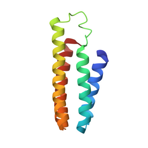
1LM3 - PubMed Abstract:
The redox potential of cytochromes sets the energy yield possible in metabolism and is also a key determinant of the rate at which redox reactions proceed. Here, the heme protein, cytochrome b(562), is used to study the in vitro evolution of redox potential within a library of variants containing the same structural archetype, the four-helix bundle. Multisite variations in the active site of cytochrome b(562) were introduced. A library of variants containing random mutations in place of R98 and R106 was created, and the redox potentials of a statistical sampling of this library were measured. This procedure was carried out for both the low- and high-potential variants of a previously studied F61X/F65X, first-generation library [Springs, S. L., Bass, S. E., and McLendon, G. L. (2000) Biochemistry 39, 6075]. The second-generation library reported here has a range of redox potentials which is greater than 40% (160 mV) of the known accessible potential among cytochromes with identical axial ligands (but different folds) and exceeds the range exhibited phylogenetically by the cytochrome c' family which internally maintains the same axial ligation and fold. A statistical analysis of the libraries examined reveals that the redox potential of WT cyt b(562) is found at the high-potential extremum of the distribution, indicating that this protein apparently evolved to differentially stabilize the reduced protein. The 2.7 A crystal structure of F61I/F65Y/R106L (low-potential variant of the second-generation library) was solved and is compared to the wild-type structure and the 2.2 A resolution structure of the F61I/F65Y variant (low-potential variant of the first-generation library). The structures indicate that charge-dipole effects are responsible for shifting the redox equilibrium toward the oxidized state in both the F61I/F65Y and F61I/F65Y/R106L variants. Specifically, a new protein dipole is introduced into the heme microenvironment as a result of the F65Y mutation, two new internal water molecules (one in hydrogen-bonding distance of Y65) are found, and in the case of F61I/F65Y/R106L (DeltaE(m) = 158 mV vs NHE), increased solvent exposure of the heme as a result of the R106L substitution is identified.
Organizational Affiliation:
Department of Chemistry, Princeton University, Princeton, New Jersey 08544, USA. ssprings@princeton.edu