1L34

HIGH-RESOLUTION STRUCTURE OF THE TEMPERATURE-SENSITIVE MUTANT OF PHAGE LYSOZYME, ARG 96 (RIGHT ARROW) HIS


Experimental Data Snapshot

  • Method: X-RAY DIFFRACTION
  • Resolution: 1.90 Å
  • R-Value Observed: 0.176 

wwPDB Validation   3D Report Full Report


This is version 2.0 of the entry. See complete history


Literature

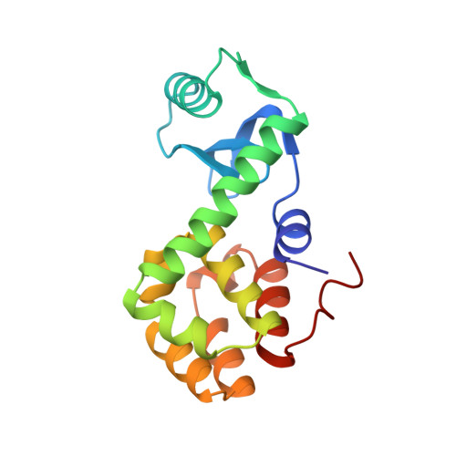
High-resolution structure of the temperature-sensitive mutant of phage lysozyme, Arg 96----His.

Weaver, L.H.Gray, T.M.Grutter, M.G.Anderson, D.E.Wozniak, J.A.Dahlquist, F.W.Matthews, B.W.

(1989) Biochemistry 28: 3793-3797

  • DOI: https://doi.org/10.1021/bi00435a025
  • Primary Citation of Related Structures:  
    1L34

  • PubMed Abstract: 

    The structure of the temperature-sensitive mutant lysozyme of bacteriophage T4 in which arginine 96 is replaced by histidine has been determined crystallographically and refined to a residual of 17.6% at 1.9-A resolution. Overall, the three-dimensional structure of the mutant protein is extremely similar to that of wild type. There are local distortions in the mutant structure suggesting that the substituted His 96 residue is under strain. This appears to be one of the major reasons for the decreased thermostability. In wild-type lysozyme the guanidinium of Arg 96 is located at the carboxy terminus of alpha-helix 82-90 and makes a pair of hydrogen bonds to two of the carbonyl groups in the last turn of the helix. The loss of this "helix dipole" interaction also appears to contribute to the destabilization. The pKa* of His 96 in the mutant lysozyme has been determined by nuclear magnetic resonance and found to be 6.8 at 10 degrees C. This relatively normal value of the histidine pKa* suggests that the protonated and unprotonated forms of the imidazole ring are perturbed equally by the protein environment or, what is equivalent, the mutant lysozyme is equally stable with either histidine species.


  • Organizational Affiliation

    Institute of Molecular Biology, University of Oregon, Eugene 97403.


Macromolecules
Find similar proteins by:  (by identity cutoff)  |  3D Structure
Entity ID: 1
MoleculeChains Sequence LengthOrganismDetailsImage
T4 LYSOZYME164Tequatrovirus T4Mutation(s): 1 
EC: 3.2.1.17
UniProt
Find proteins for P00720 (Enterobacteria phage T4)
Explore P00720 
Go to UniProtKB:  P00720
Entity Groups  
Sequence Clusters30% Identity50% Identity70% Identity90% Identity95% Identity100% Identity
UniProt GroupP00720
Sequence Annotations
Expand
  • Reference Sequence
Experimental Data & Validation

Experimental Data

  • Method: X-RAY DIFFRACTION
  • Resolution: 1.90 Å
  • R-Value Observed: 0.176 
  • Space Group: P 32 2 1
Unit Cell:
Length ( Å )Angle ( ˚ )
a = 60.8α = 90
b = 60.8β = 90
c = 96.8γ = 120
Software Package:
Software NamePurpose
TNTrefinement
AGROVATA / ROTAVATAdata scaling

Structure Validation

View Full Validation Report


Entry History 

Deposition Data

Revision History  (Full details and data files)

  • Version 1.0: 1990-01-15
    Type: Initial release
  • Version 1.1: 2008-03-24
    Changes: Version format compliance
  • Version 1.2: 2011-07-13
    Changes: Advisory, Version format compliance
  • Version 1.3: 2017-11-29
    Changes: Derived calculations, Other
  • Version 1.4: 2020-07-22
    Changes: Advisory, Data collection, Database references, Other, Refinement description
  • Version 2.0: 2022-11-23
    Type: Remediation
    Changes: Advisory, Atomic model, Data collection, Database references, Other, Source and taxonomy