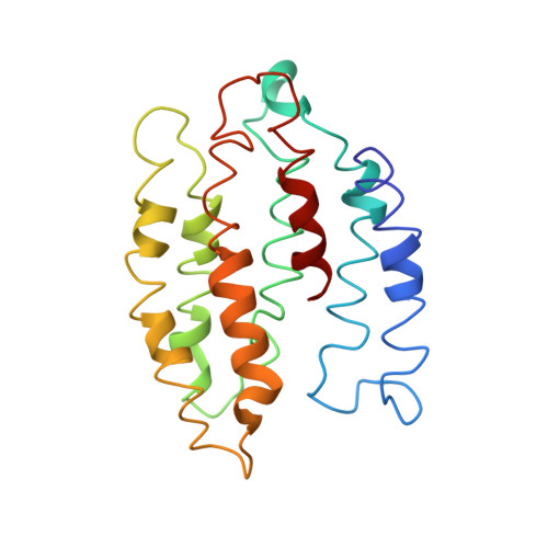
Assembly of a polytopic membrane protein structure from the solution structures of overlapping peptide fragments of bacteriorhodopsin.
Katragadda, M., Alderfer, J.L., Yeagle, P.L.(2001) Biophys J 81: 1029-1036
- PubMed: 11463644
- DOI: https://doi.org/10.1016/S0006-3495(01)75760-8
- Primary Citation of Related Structures:
1L0M - PubMed Abstract:
Three-dimensional structures of only a handful of membrane proteins have been solved, in contrast to the thousands of structures of water-soluble proteins. Difficulties in crystallization have inhibited the determination of the three-dimensional structure of membrane proteins by x-ray crystallography and have spotlighted the critical need for alternative approaches to membrane protein structure. A new approach to the three-dimensional structure of membrane proteins has been developed and tested on the integral membrane protein, bacteriorhodopsin, the crystal structure of which had previously been determined. An overlapping series of 13 peptides, spanning the entire sequence of bacteriorhodopsin, was synthesized, and the structures of these peptides were determined by NMR in dimethylsulfoxide solution. These structures were assembled into a three-dimensional construct by superimposing the overlapping sequences at the ends of each peptide. Onto this construct were written all the distance and angle constraints obtained from the individual solution structures along with a limited number of experimental inter-helical distance constraints, and the construct was subjected to simulated annealing. A three-dimensional structure, determined exclusively by the experimental constraints, emerged that was similar to the crystal structure of this protein. This result suggests an alternative approach to the acquisition of structural information for membrane proteins consisting of helical bundles.
Organizational Affiliation:
Department of Molecular and Cell Biology, University of Connecticut, Storrs, Connecticut 06269, USA.